


- Whereas tumors distort adrenal shape, the normal triangular configuration of the gland is usually preserved in cortical hemorrhage.
- It is frequently discovered as an incidental finding at US performed for other reasons.
- The pathogenesis is unclear, but such hemorrhage has been associated with birth trauma (especially in infants large for their gestational age, such as those born of diabetic mothers), overwhelming septicemia, and hypoxia.
- Idiopathic neonatal adrenal hemorrhage is most often asymptomatic but may manifest as a unilateral flank mass, hypovolemic shock, anemia, or jaundice.
- Although it has been observed in utero, it usually occurs around birth.
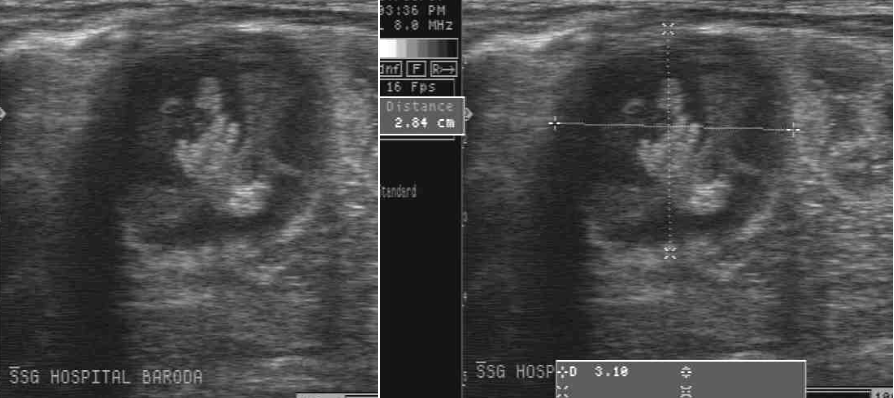
- Diagnosis of uncomplicated neonatal hemorrhage is based on sequential sonographic examination, which initially demonstrates a solid appearing mass followed by liquefaction and regression in size.
- CT and MR imaging demonstrate characteristic findings but are usually not required, except when the mass remains solid in appearance over time or enlarges or coexisting neuroblastoma needs to be ruled out.
- Usually the hemorrhage resolves completely, but may calcify at its periphery as seen incidentally on plain abdominal radiographs or CT.
Other causes of AH include-
1.Abdominal injuries
2.Rt. AH asso: with prior liver transplantation
3.Lt. AH asso: with renal vein thrombosis
4.Benign hemorrhagic cysts seen in Beckwith-Wiedemann Syndrome
DDs of neonatal adrenal lesions also include
DDs of neonatal adrenal lesions also include
1.Neuroblastoma
2.Congenital Adrenal Hyperplasia
3.Adrenal cortical tumors
4.Storage diseases & diffuse adrenal diseases