


Imaging finding:
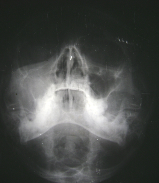
Radiograph showing opacification of maxillary sinus
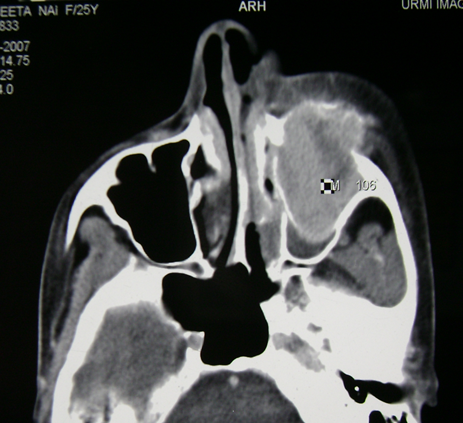
CT- Expansile hyperenchancing lesion Is noted in the left maxillary sinus. It is causing expansion of the maxillary sinus.
Histology findings.
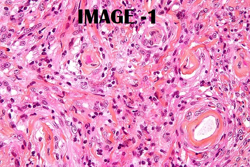
Image 1 - Micrograph of a meningioma showing the characteristic whorling, HPS stain. Image courtesy – Wikimedia commons
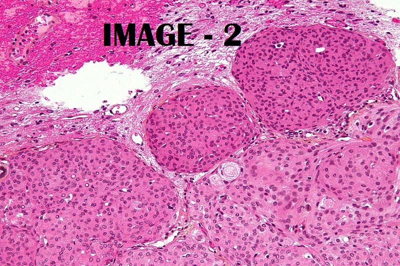
Image 2- Micrograph of a meningioma with brain invasion (WHO Grade II); the tumour (bottom/right of image) has the typical "pushing border" invasion into the cerebral cortex (top/left of image), HPS stain. Image courtesy – Wikimedia commons
Diagnosis- Primary extracranial Meningioma
Discussion
· Meningioma is the most common nonglial intracranial neoplasm, arising from meningocytes (arachnoid cap cells) of arachnoid granulation tissue.
· Approximately 6-17% of all meningiomas can be found in extracranial or extraspinal sites.
The classification system of Hoye delineates the major etiologies of the tumor and its development:
A. Extracranial extension of an intracranial meningioma (secondary).
B. Extracranial extensions of a meningioma arising in a neural foramen (primary).
C. Ectopic, without any connection either to a foramen of a cranial nerve or to intracranial structures (primary).
D. Extracranial metastasis of a malignant intracranial meningioma (secondary).
Our case belong to category 3 ie Primary Extracranial Meningioma
· PEM are rare, mostly in the head and neck region, such as the skull bones, scalp, nose, orbit,paranasal sinuses, parotid gland, pharynx, mandible & middle ear, the lung, mediastinum, skin of the finger and eyelid, retroperitoneum and the thigh muscle.
· In most cases, th,eir morphological features are similar to those of a meningioma of the central nervous system.
· It has been suggested that ectopic meningiomas arise from ectopic arachnoid cells or from mesenchymal cells or Schwann cells that have differentiated into meningothelial cells.
· The clinical presentation of PEM can be extremely subtle. Localizing signs or symptoms usually become apparent only when the tumor reaches significant size. Manifestations are related to mass effect at the site and neurological dysfunctions due to intimate involvement of cranial nerves.
· Rarely, ectopic meningiomas arise through metastatic spread, in the lung, liver, bone and lymph nodes, and are often seen with malignant variants.
DDs
- Tumors of epithelial origin such as squamous cell carcinoma,
- Neurogenic origin such as esthesioneuroblastoma,
- Odontogenic tissues such as sarcoma and ameloblastoma,
- Vascular origin such as angiofibroma and
- Haematological such as lymphoma
The histopathologic report came as a meningioma.