


IMAGING
- Osseous fragility,blue sclera,otosclerosis,Dentinogenesis imperfecta
- Postnatal x-ray: Obtain skull, chest, long bones, and pelvis x-ray films.
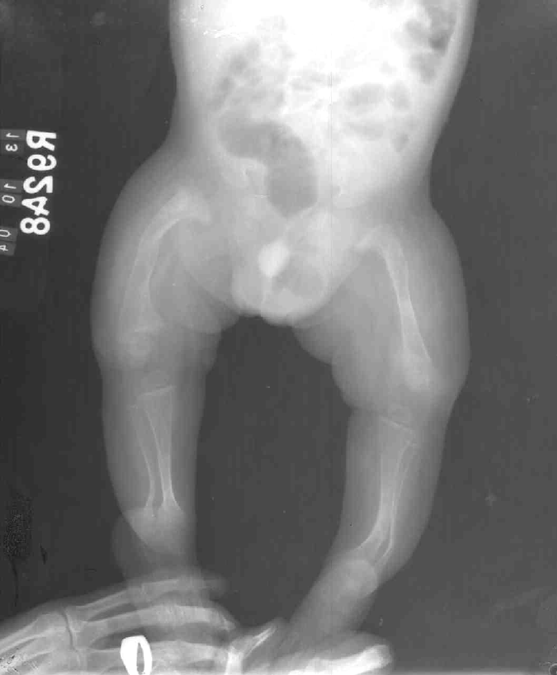
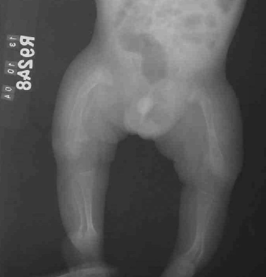
- Type I
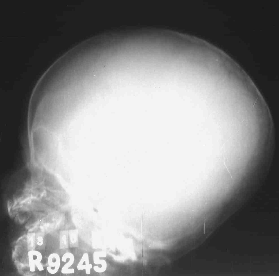
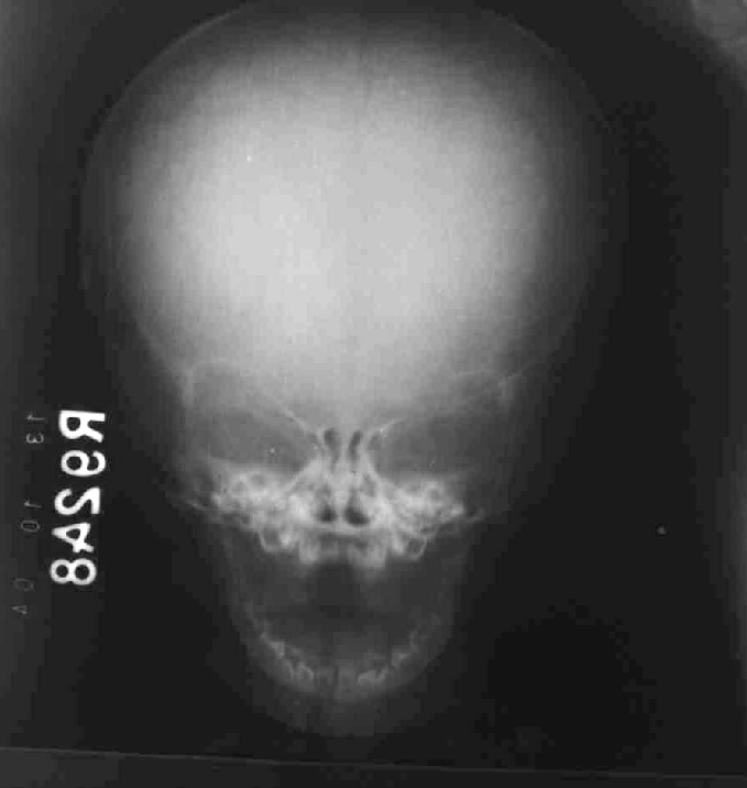
- Tam O'Shanter skull - Flattening in vertical axis and widening in transverse axis
- Thin bones
- Multiple wormian bones
- Fractures with deformities
- Osteopenia
- Platyspondylia
-
- Tam O'Shanter skull - Flattening in vertical axis and widening in transverse axis
- Type I
- A: Dentinogenesis imperfecta is absent.
- B: Dentinogenesis imperfecta is present.
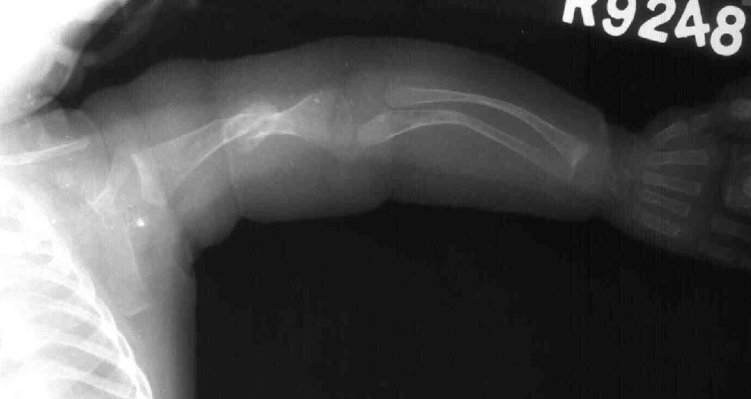
- Type II
- Beaded ribs
- Broad bones
- Fractures with deformities
- Osteopenia
- Platyspondylia
- Blue sclera may be present
- In utero fractures are present in 100% cases .
- Beaded ribs
- Type III
- Cystic metaphyses (popcorn appearance)
- Normal or broad bones early on; thin bones later
- Fractures with deformities
- Osteopenia
- Sclera of variable hue
-
- Cystic metaphyses (popcorn appearance)
- Type IV
- Thin bones
- Fractures with deformities
- Osteopenia
- Thin bones
- Prenatal ultrasound
- Prenatal ultrasound can be used to detect limb length abnormalities at 15-16 weeks’ gestation.
- Limb abnormality can be detected by 15-18 weeks’ gestation. Mild forms may have normal sonogram findings.
- Features
- Supervisualization of intracranial contents caused by decreased mineralization of calvarium (also calvarial compressibility)
- Long bones bowing, decrease in length (especially the femur)
- Multiple rib fractures
- Supervisualization of intracranial contents caused by decreased mineralization of calvarium (also calvarial compressibility)
-