


Silicosis is a fibronodular lung disease caused by inhalation of dust containing crystalline silica (alpha-quartz or silicon dioxide), which is distributed widely, or its polymorphs (tridymite or cristobalite), which are distributed less widely. Associated occupations include mining or tunneling, quarrying, drilling, crushing stone, chipping, grinding, sandblasting, grinding or polishing in pottery and foundry work, cement manufacturing, construction industry, cutting or manufacturing heat-resistant bricks.
Small (£1 mm) particles are more dangerous because they are more likely to be deposited distally in the respiratory bronchioles, alveolar ducts, and alveoli. The surface of these particles generates silicon-based radicals that lead to the production of hydroxyl, hydrogen peroxide, and other oxygen radicals that damage cell membranes by lipid peroxidation and inactivate essential cell proteins. Alveolar macrophages ingest the particles, become activated, and release cytokines. The ensuing inflammation damages resident cells and the extracellular matrix.
As the disease progresses, airflow limitation occurs, manifested by dyspnea and cough. Eventually, cor pulmonale and respiratory failure develop. An increased incidence of mycobacterial diseases is reported in patients with silicosis. Three types of silicosis are seen:
1.Simple chronic silicosis
2.Accelerated silicosis
3.Acute silicosis
Progressive massive fibrosis may occur in simple or accelerated silicosis, but is more common in the accelerated form. Progressive massive fibrosis results from severe scarring and leads to obliteration of normal lung structures.
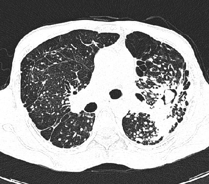
The radiological findings in Silicosis and CWP are similar and are characterized by development of multiple nodules predominantly in the upper lobes , more so posteriorly. As the disease progresses these nodules coalesce to from large nodular opacities (diam > 1cm), asstd with clinical deterioration of the patient. Large nodular opacities can form large conglomerate of masses or PMF and distort lung architecture and is accompanied by development of cicatrical emphysema.CT/HRCT shows fibrosis with emphysema. CT can monitor disease progression and evaluate for potentially curable superimposed lung disease such as TB or lung cancer.