


Von Hippel-Lindau syndrome (VHL) is an autosomal dominant, inherited, neurocutaneous dysplasia complex with an 80-100% penetrance and variable delayed expressivity. Sex distributions are equal, and 20% of cases are familial.The gene has been located on chromosome bands 3p25-26.
Imaging
Imaging
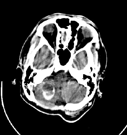
- Hemangioblastomas of the CNS are demonstrated as cystic lesions with a 3- to 15-mm mural nodule in 75% of patients. They are demonstrated as an enhancing lesion with multiple cystic areas in 15% of patients and as an enhancing solid mass in 10%.
- Hemangioblastomas usually do not become calcified, and this is a finding that helps in differentiating these lesions from cystic astrocytomas (which are calcified in 25% of patients). Typical pilocytic cystic astrocytomas also occur in patients much younger than most patients with hemangioblastomas.
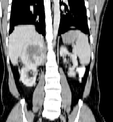
- CT has a low sensitivity in the detection of renal cell carcinoma associated with VHL because of its inability to reliably differentiate cystic renal cell carcinomas, cancers within a cyst, and atypical cysts.
Location
The cerebellum is most commonly involved, followed by the medulla, the spinal cord, and even the spinal nerve roots. Supratentorial hemangioblastomas are rare.
Features
The cerebellum is most commonly involved, followed by the medulla, the spinal cord, and even the spinal nerve roots. Supratentorial hemangioblastomas are rare.
Features
VHL is characterized by a predisposition to bilateral and multicentric retinal angiomas, central nervous system (CNS) hemangioblastomas; renal cell carcinomas; pheochromocytomas; islet cell tumors of the pancreas; endolymphatic sac tumors; and renal, pancreatic, and epididymal cysts. CNS hemangioblastoma (Lindau tumor) is the most commonly recognized manifestation of VHL and occurs in 40% of patients
Criteria for the diagnosis of VHL include the following:
(1) more than 1 hemangioblastoma in the CNS
(2) 1 CNS hemangioblastoma and visceral manifestations of VHL
(3) 1 manifestation and a known family history of VHL.
(1) more than 1 hemangioblastoma in the CNS
(2) 1 CNS hemangioblastoma and visceral manifestations of VHL
(3) 1 manifestation and a known family history of VHL.