


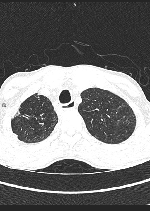
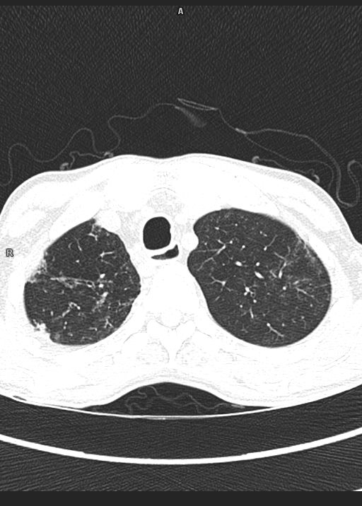
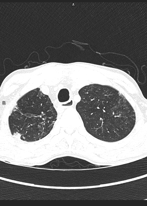
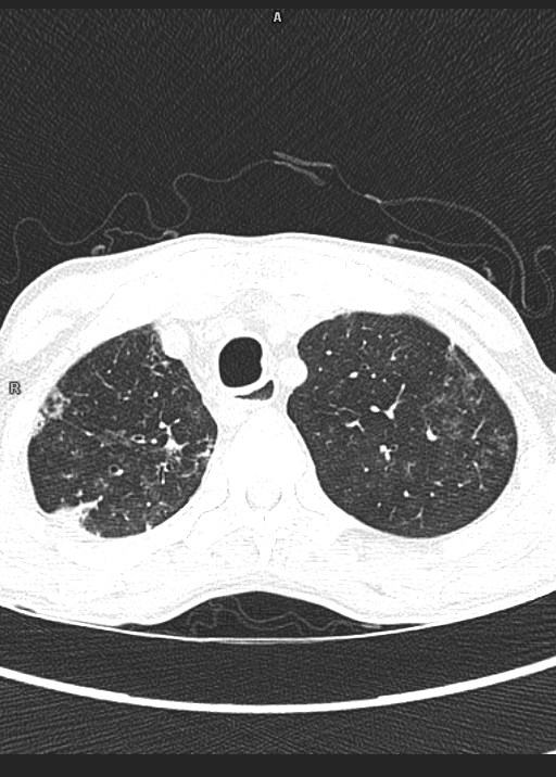


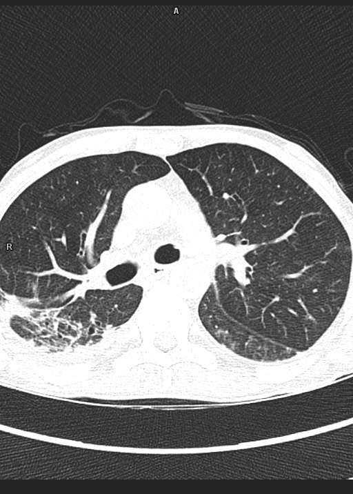
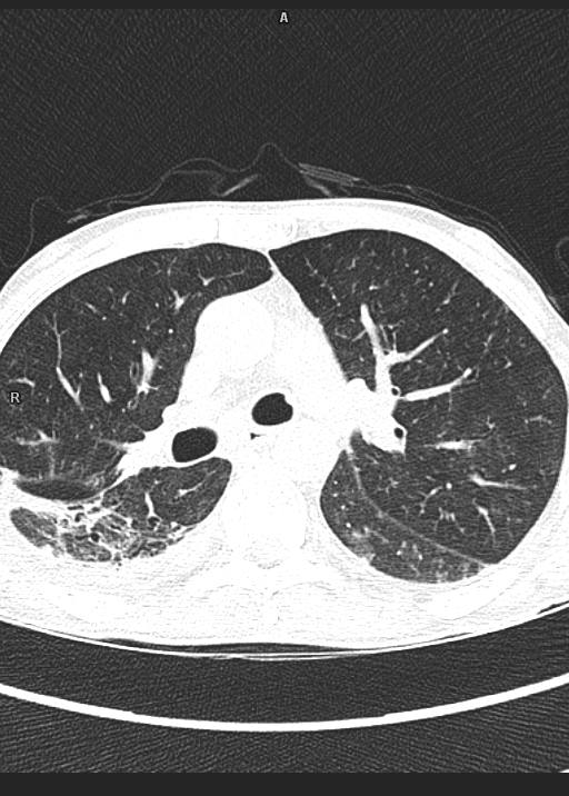
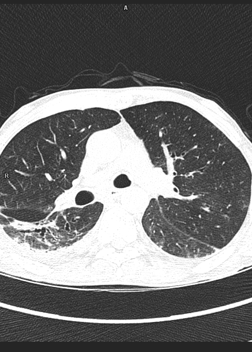
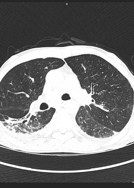
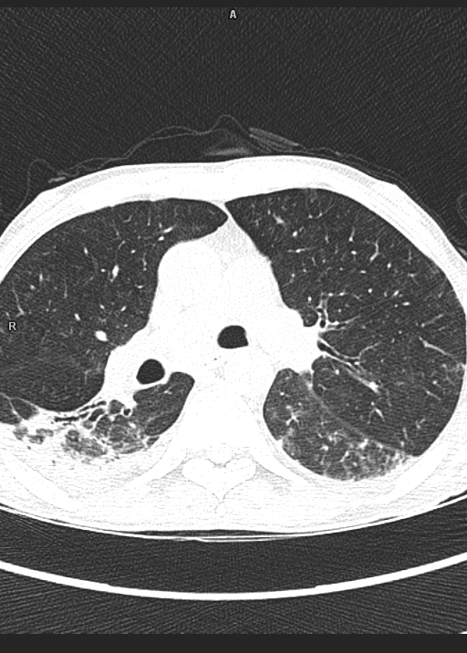


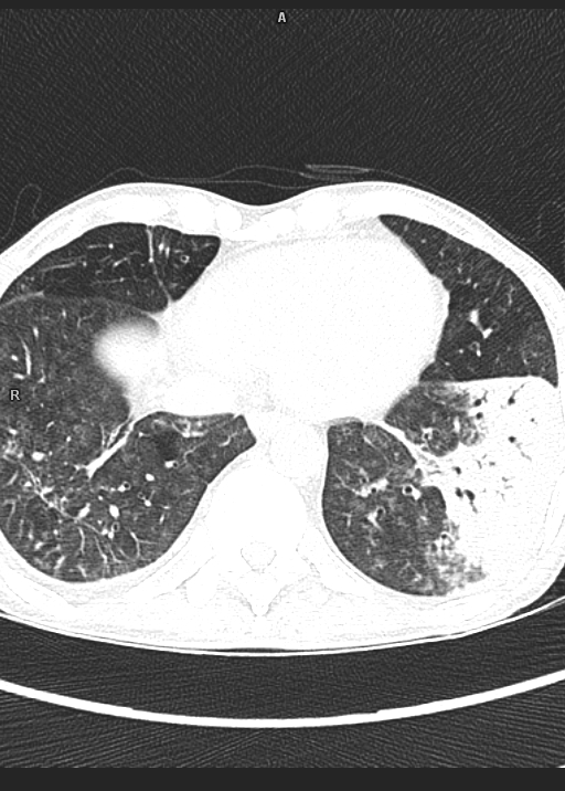
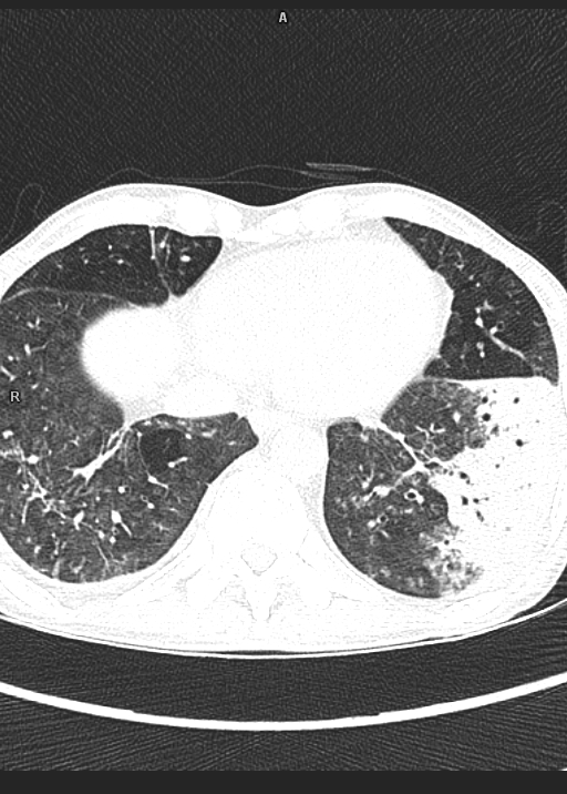
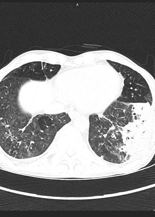
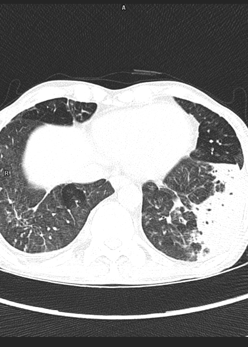
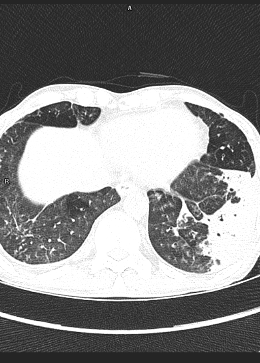
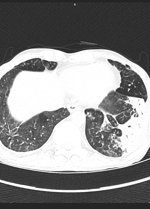
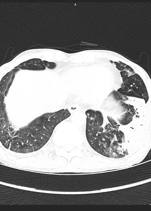
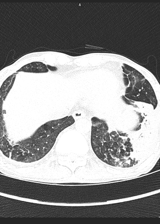
Study reveals,
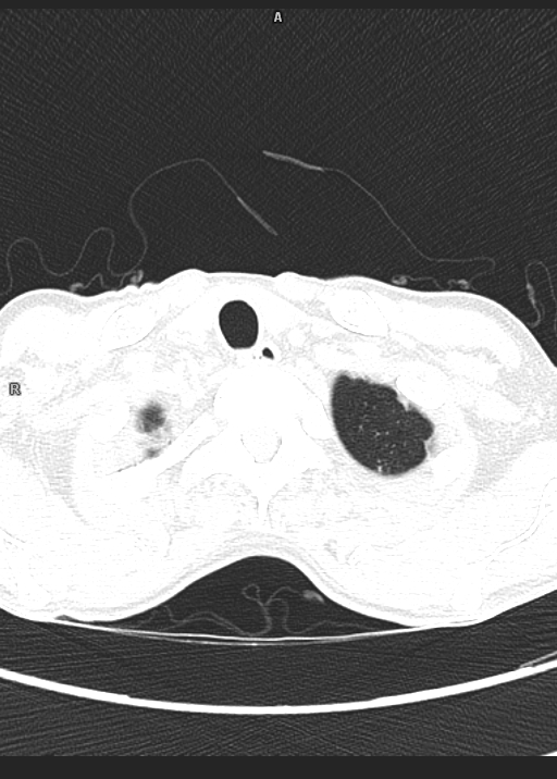
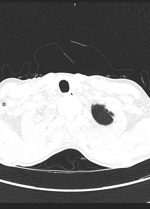
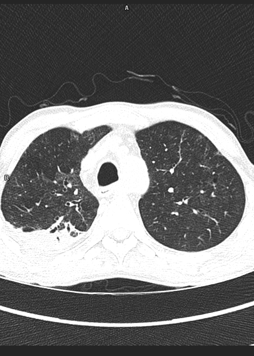
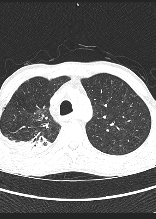
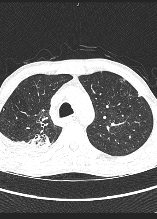
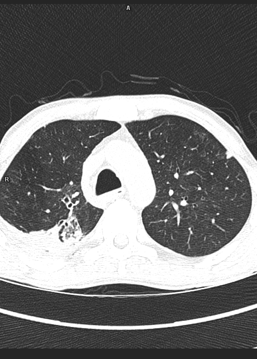
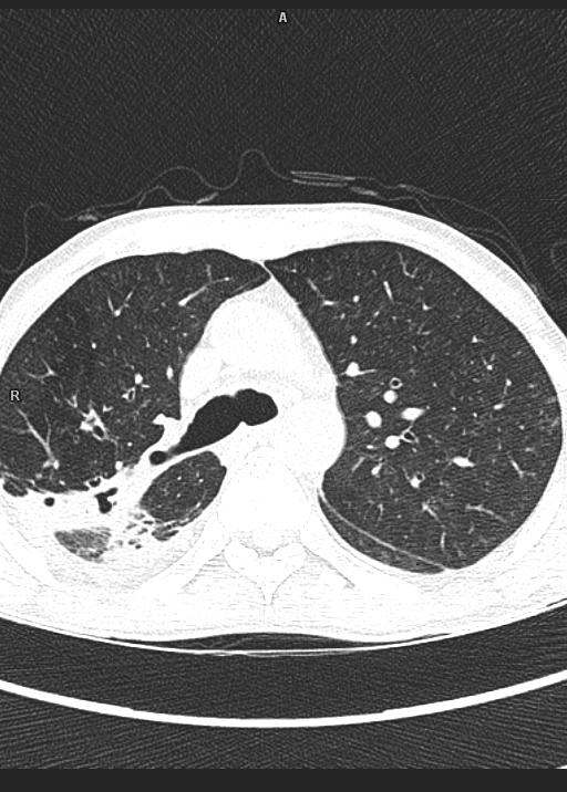
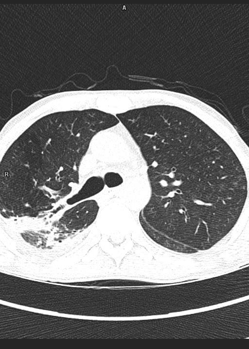
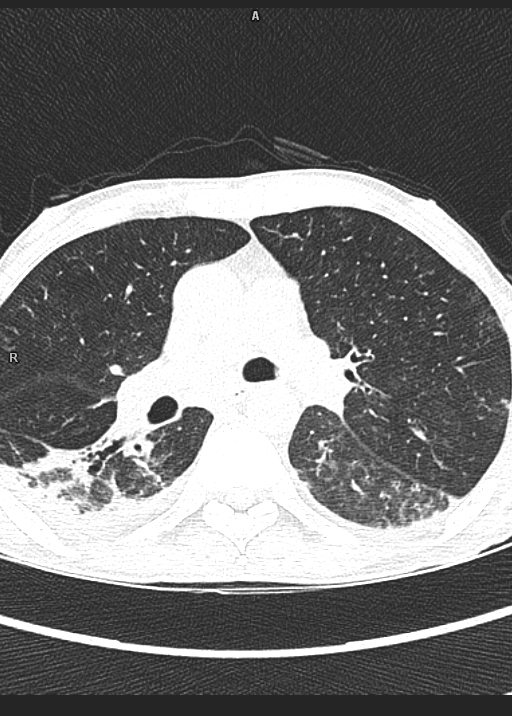
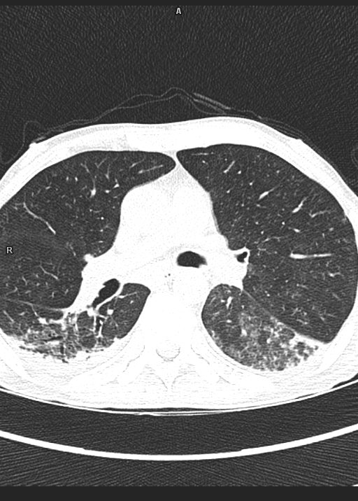
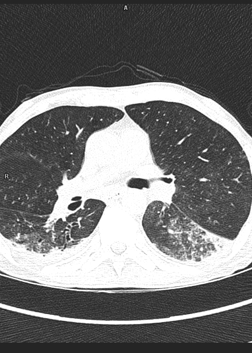
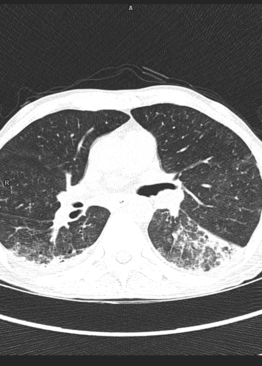
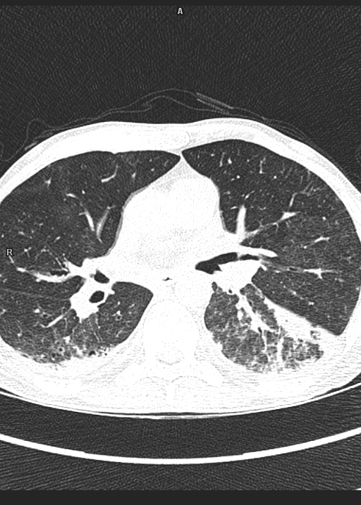
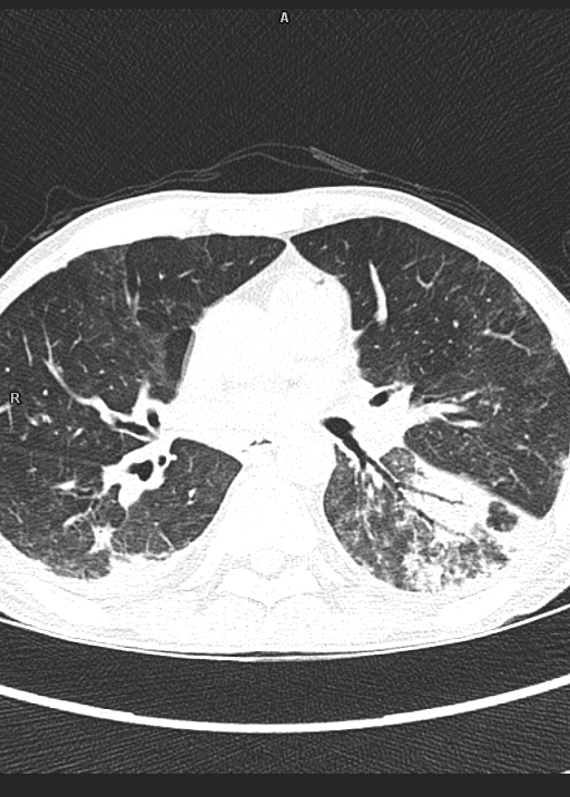
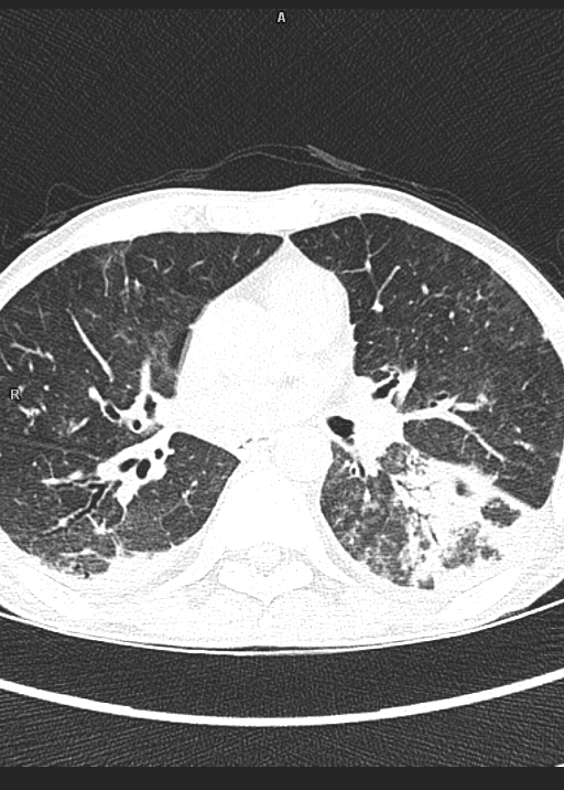
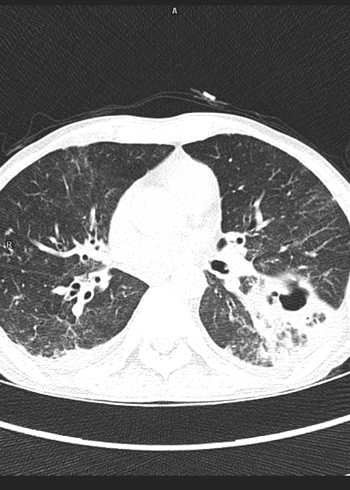
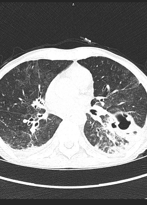
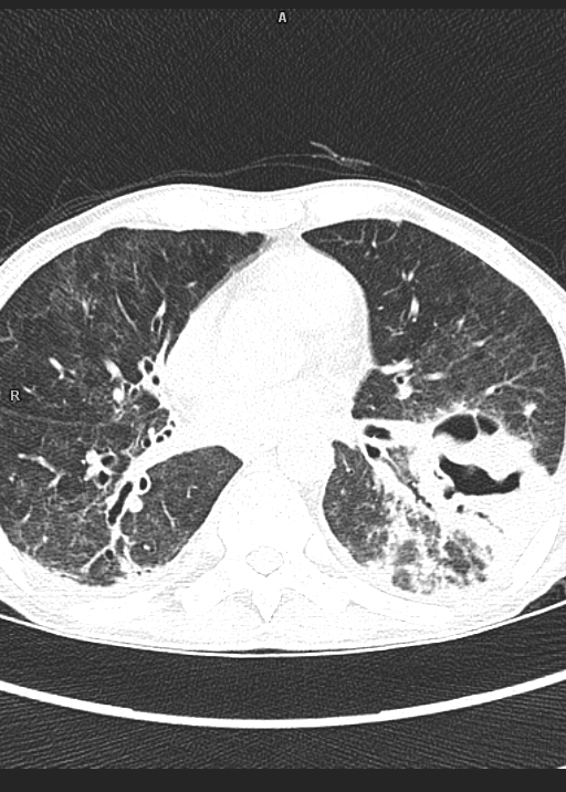
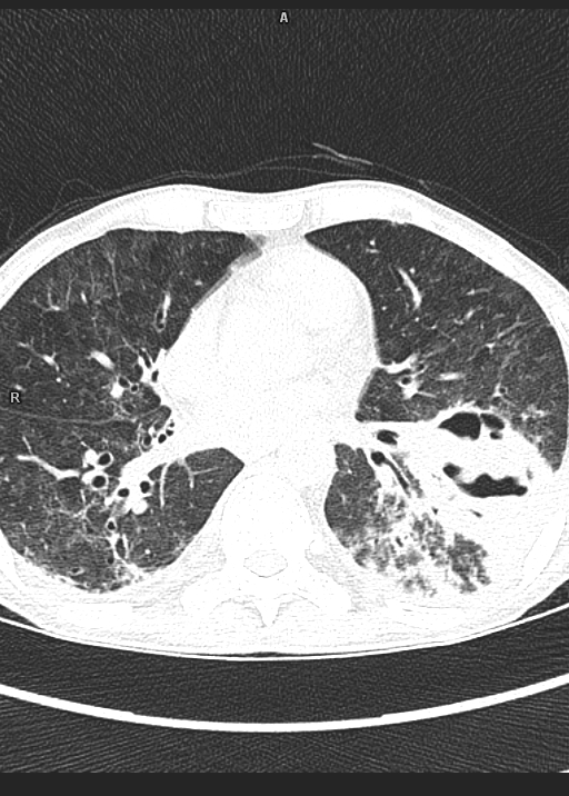
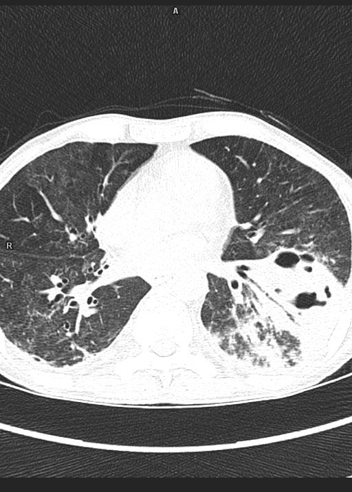
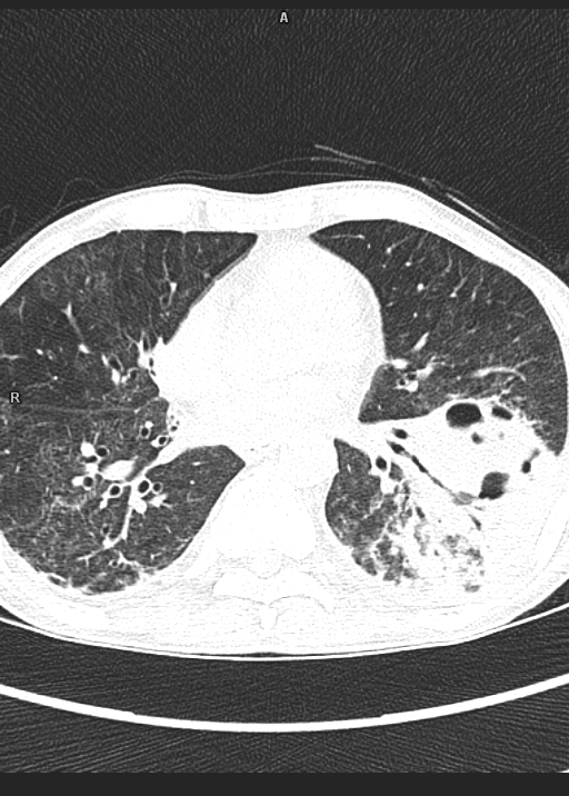
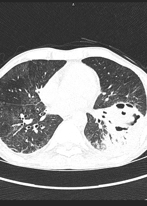
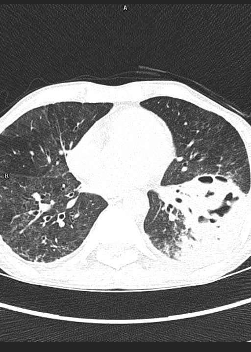
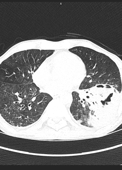
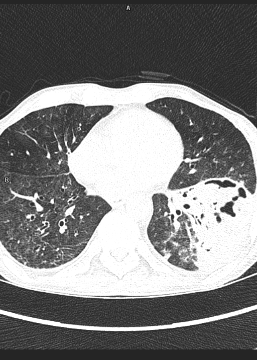
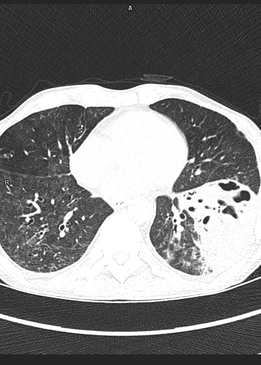
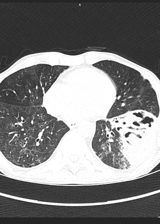
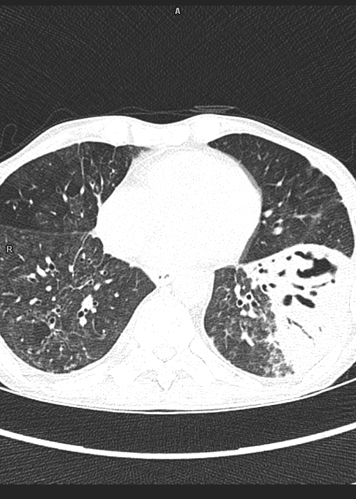
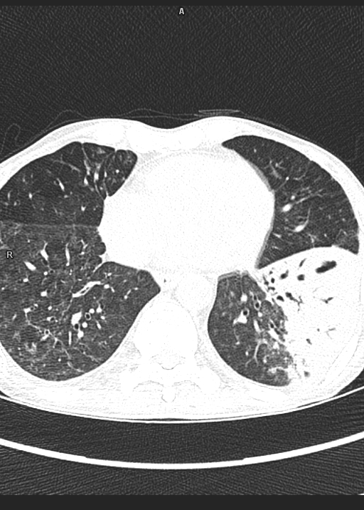
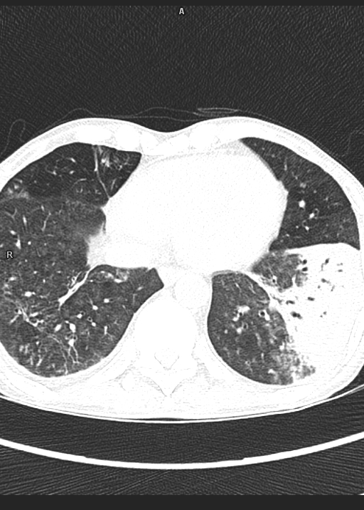
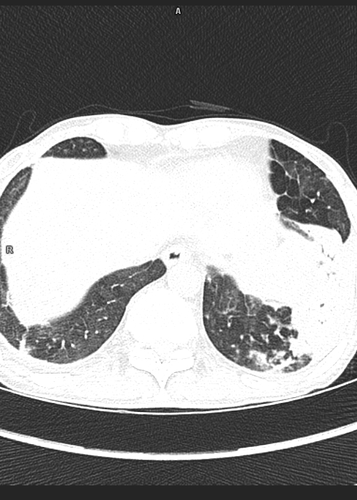
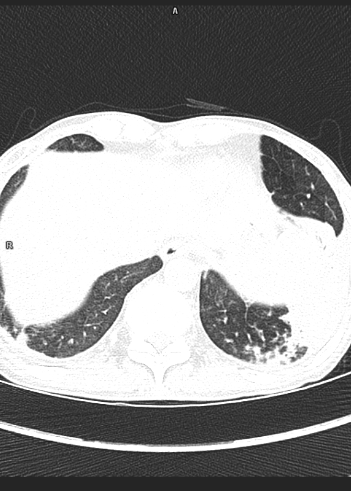
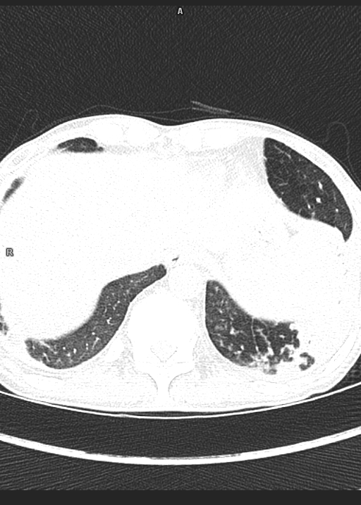
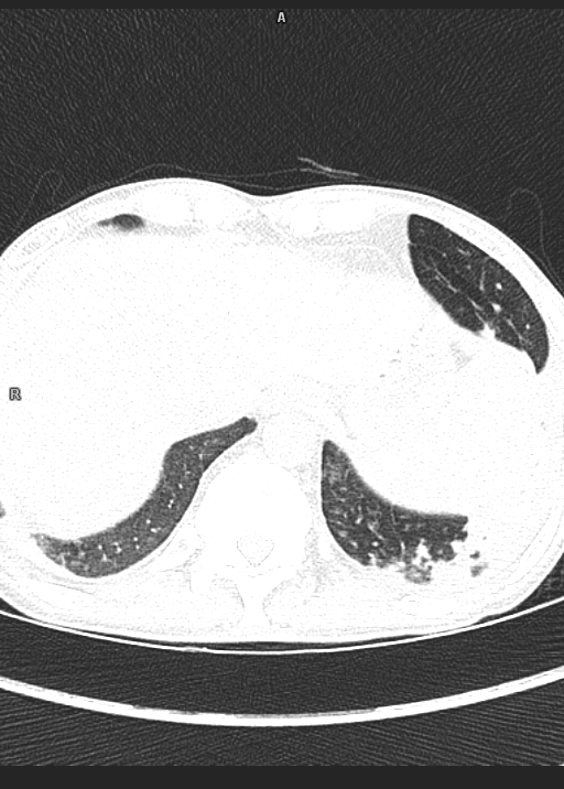
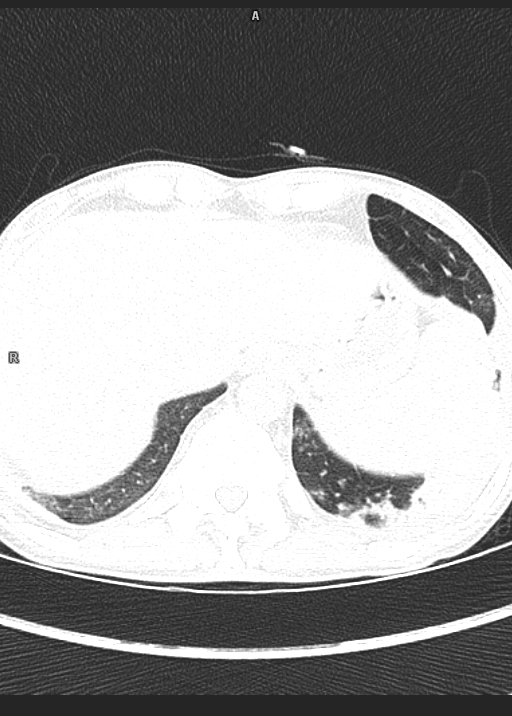
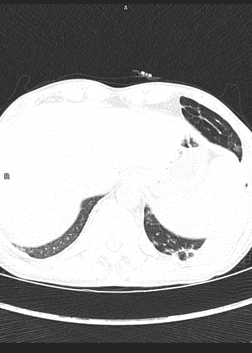
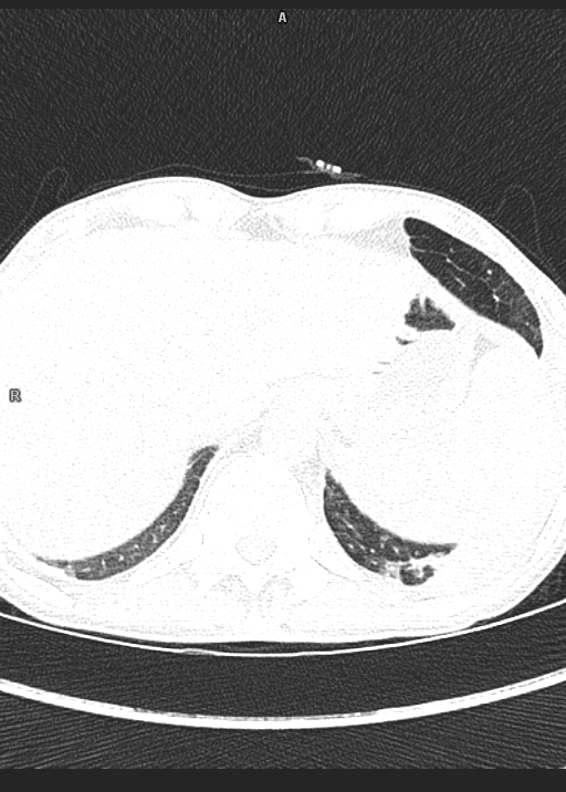
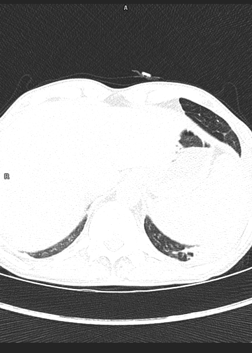
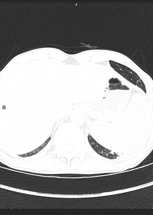
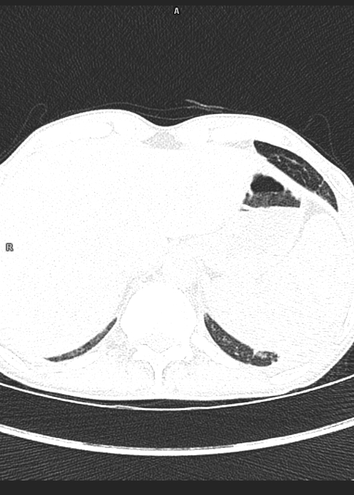
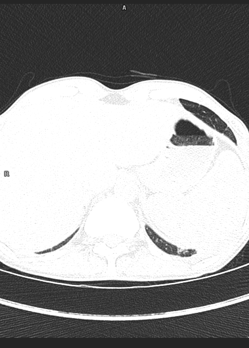
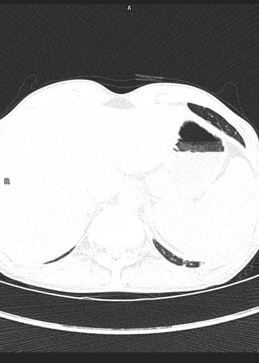
· Area of consolidation with adjacent areas of with areas of break down noted involving left lower lobe with cavity formation of size 3.8x4.3x3.2cm.
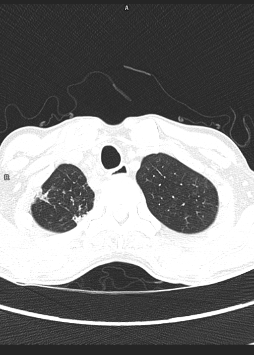
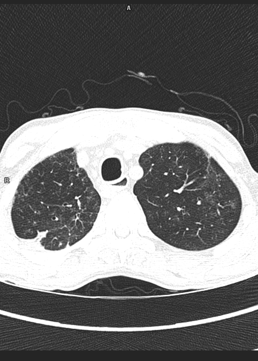
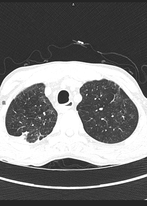
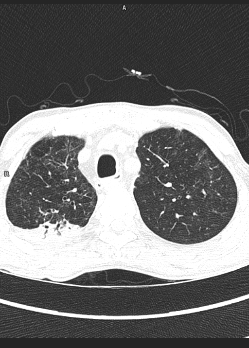
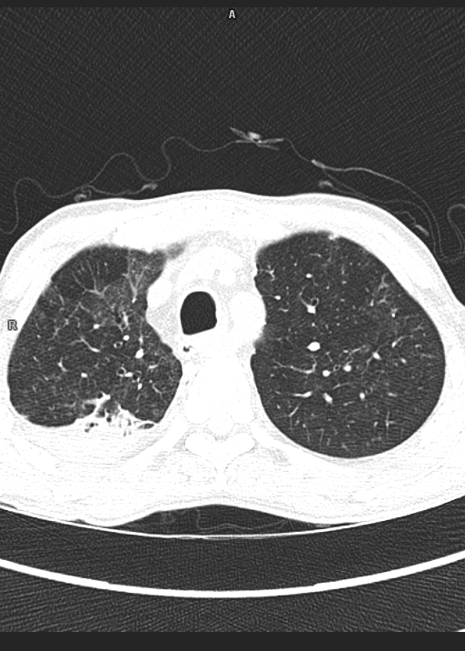
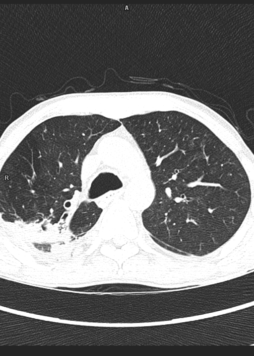
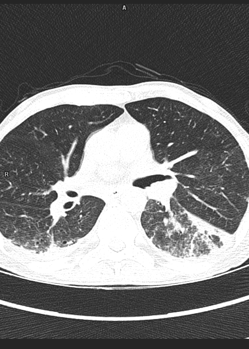
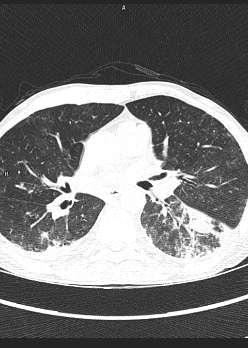
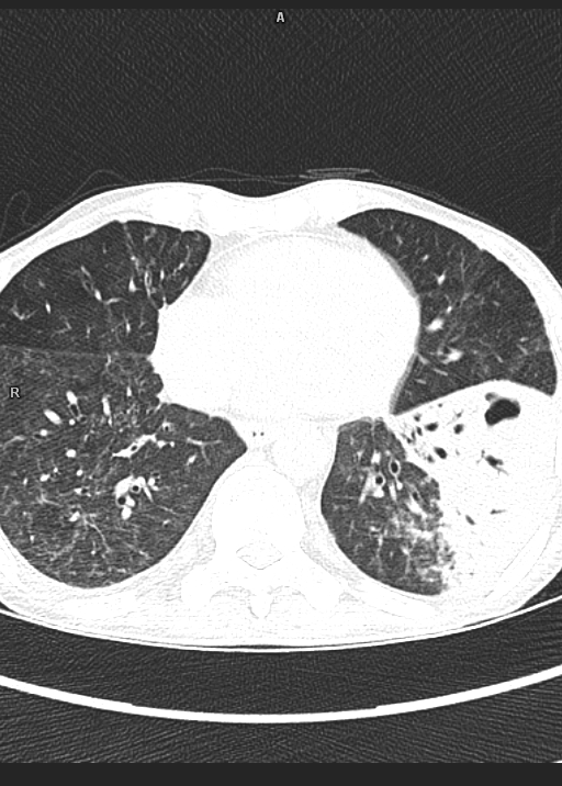
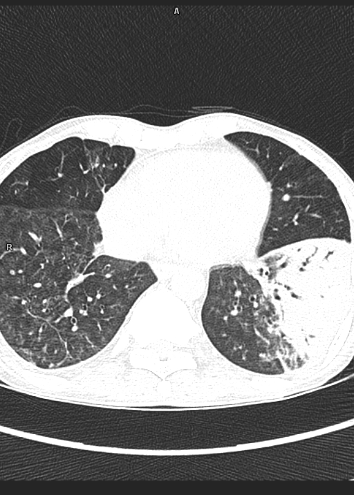
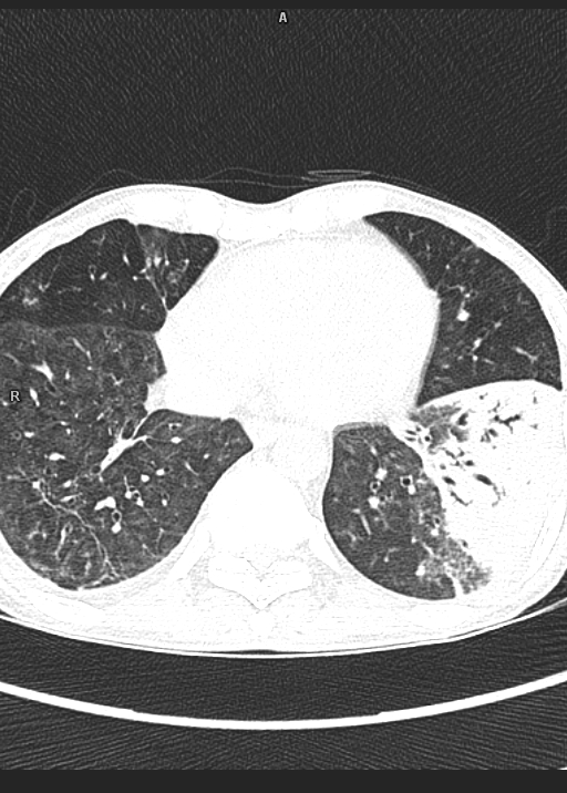
· Centrilobular nodules with adjacent ground glass opacities are seen in bilateral lower lobes.
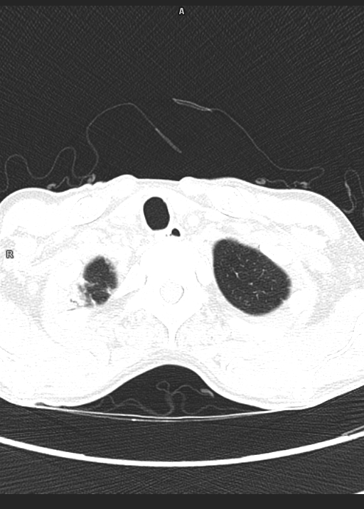
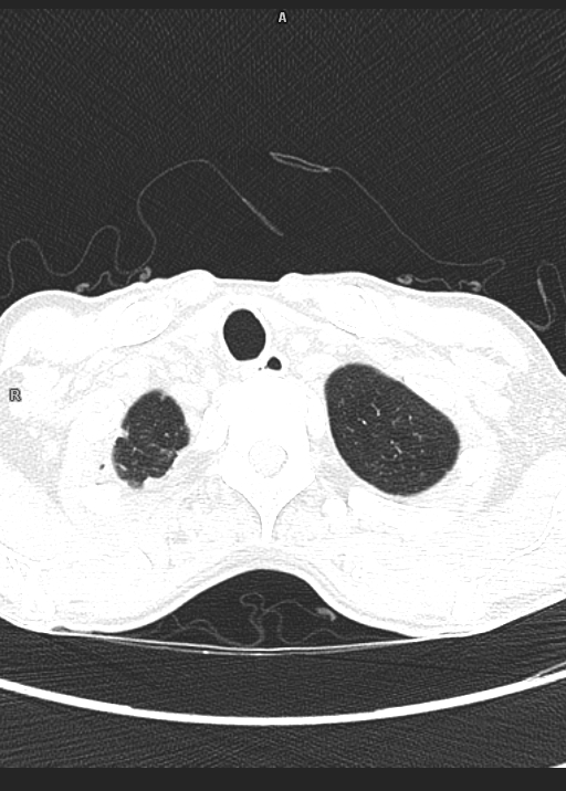
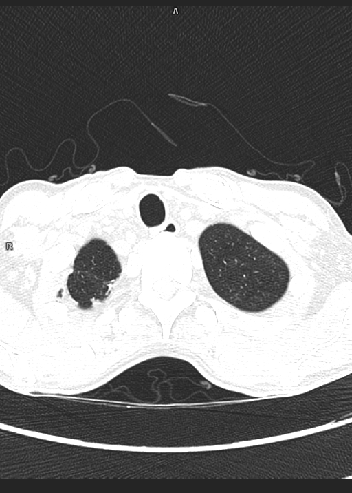
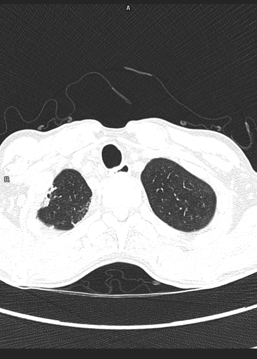
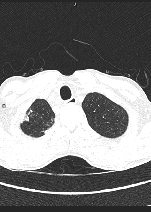
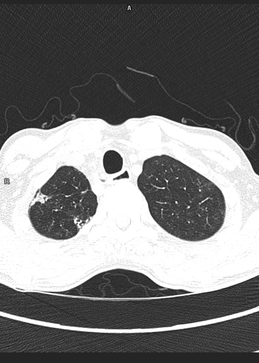
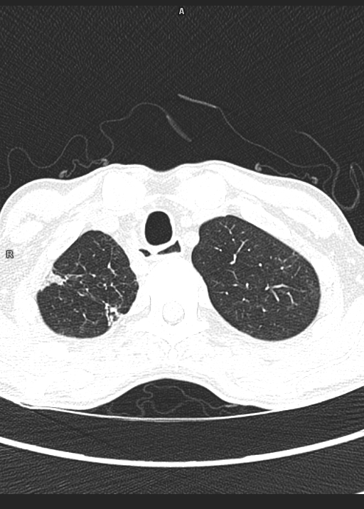
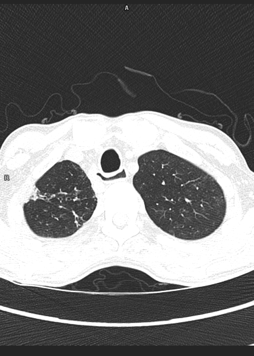
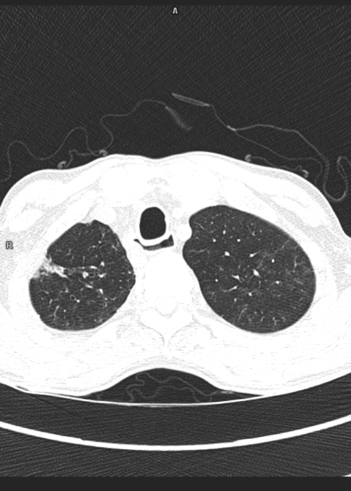
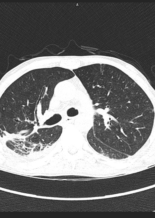
· Fibrobronchiectatic changes with fibrotic strands are noted involving posterior segment of right upper lone and apical segment of right lower lobe with severe volume loss/collapse.
· Multiple areas of fibrotic strands noted involving right upper lobe, lingular segment, and lateral basal segment of right lower lobe.
· Areas of nodular pleural thickening are noted along right apical lobe and right chest wall.
· Mosaic attenuation of right lung parenchyma is noted.
· Trachea and major bronchi are normal.
· Few enlarged lymph nodes are noted in pre/para tracheal and subcarinal region largest measuring 1.1x0.8cm in para-tracheal region.
· Cardiac chambers are normal in size.
· Mediastinal vasculature appears normal.
· Mild left sided pleural effusion is noted.
· No pericardial effusion is noted.
· Visualized bones show degenerative changes in the form of marginal osteophytes and end plate changes.
Impression:
· Areas of consolidations with adjacent areas of break down involving left lower lobe with cavity formation as described.
· Centrilobular nodules with adjacent ground glass opacities in bilateral lower lobes.
· Left sided pleural effusion.
Above features likely s/o acute on chronic infective etiology