


Clinical profile: c/o pain in epigastric region since 4 days .USG s/o bulky and heterogenous pancreas s/o acute pancreatitis and mild ascites present .guarding , tenderness present .past h/o o/c/o intestinal obstruction 10 yrs back.
Study reveals,
· Ryle’s tube noted in situ.
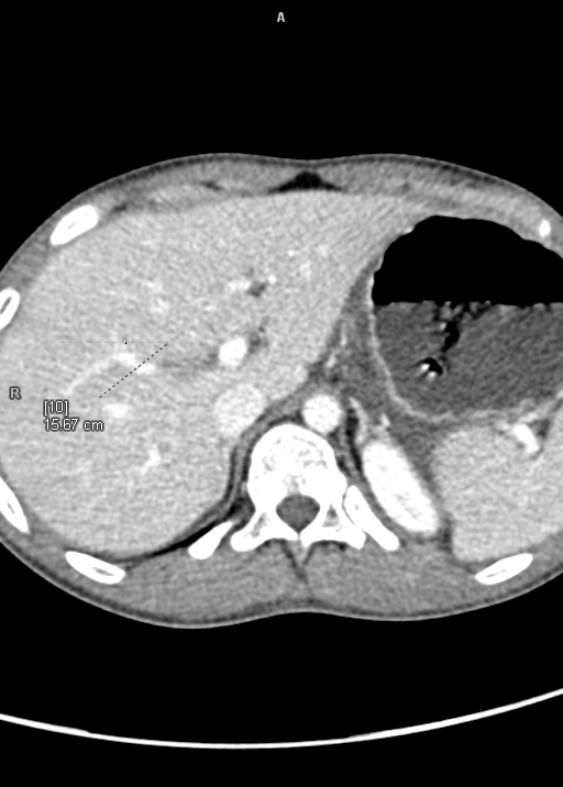
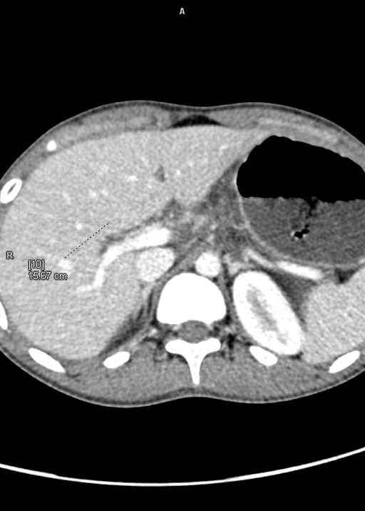
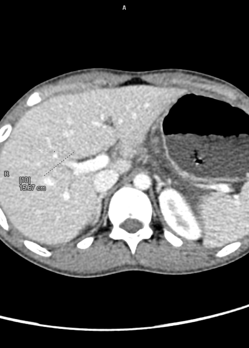
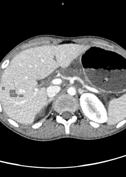
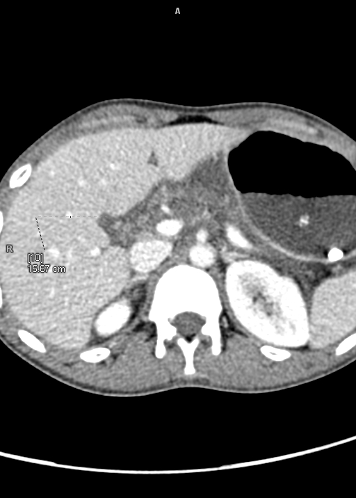
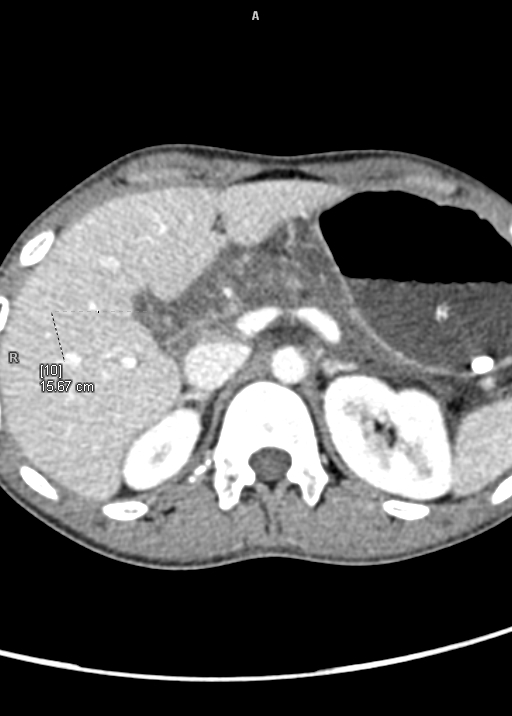
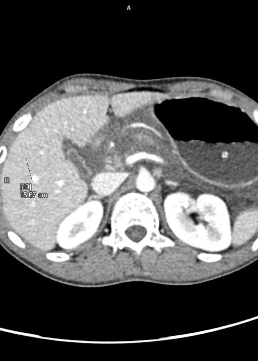
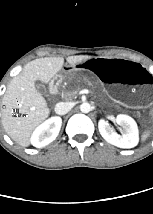
· Pancreas shows of extensive non enhancing areas involving body and tail region on contrast images with intra-pancreatic and peri-pancreatic fluid collection s/o necrosis is noted. Extensive peri-pancreatic fat stranding is noted.
· Head of the pancreas appears mildly bulky and shows homogenous contrast enhancement. Peri-pancreatic fluid collection is noted. MPD is not visualized. No e/o calcifications noted within it.
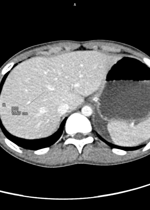
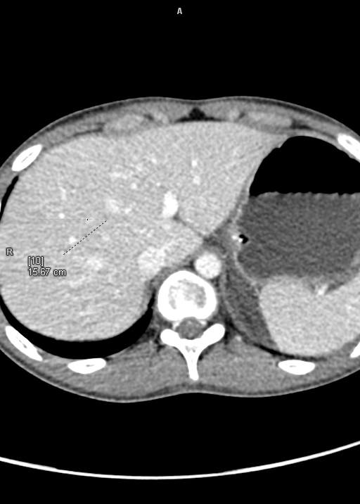
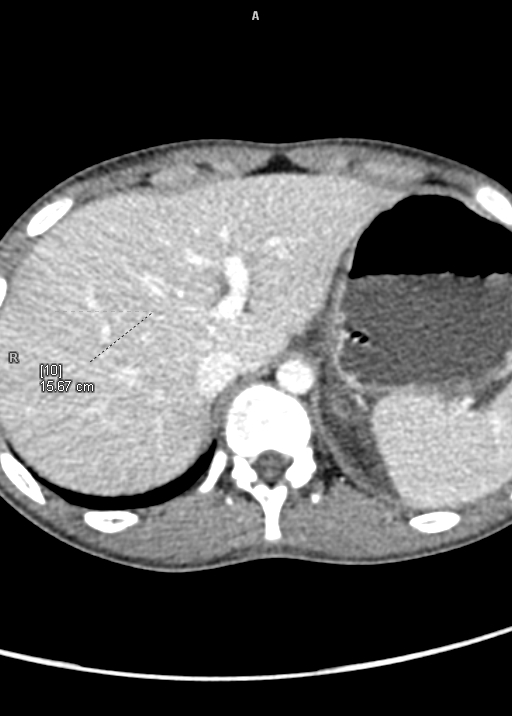
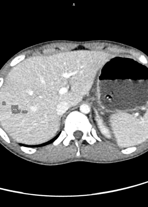
· Liver is normal in size shows normal contrast enhancement. Hypodense filling defect is noted involving segmental branch of right portal vein of segment VII branch of s/o thrombosis. Rest of the portal vein and its branches show normal contrast opacification.
· Gall bladder is distended with normal wall thickness. CBD appears normal.
· Spleen is normal in size. It shows normal contrast enhancement.
· Aorta, IVC, portal vein, SV, SMV and SMA appears normal.
· Both adrenals are normal.
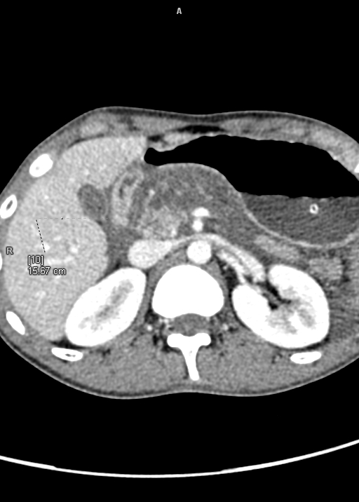
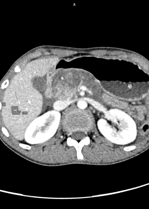
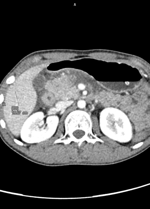
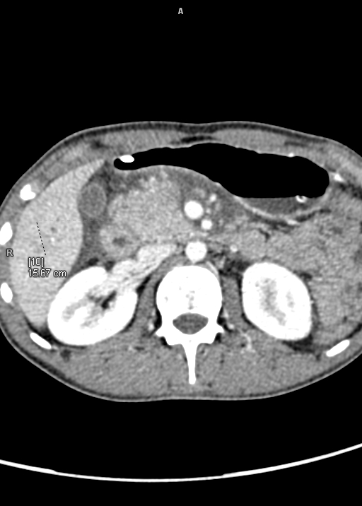
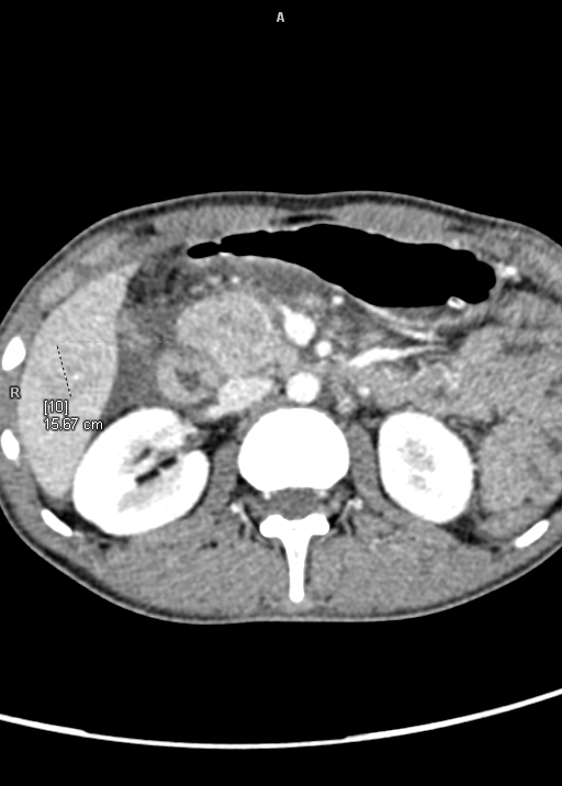
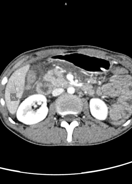
· Both kidneys appear normal in size and shows prompt nephrogram and good excretion of contrast. Both ureters appear normal in course and caliber.
· Urinary bladder appears partially distended, Foley’s bulb noted in situ.
· Prostate appears normal.
· Few enlarged peri-pancreatic, pre/para aortic lymph nodes are noted largest measuring 1.6x0.8cm.
· Moderate free fluid is noted in abdomen and pelvis.
· Visualised bowel loops appear normal. Ileoceacal junction appears normal.
· Few fibrotic strands are noted in right lower lobe..
· Visualised bones appear normal.
Impression:
· F/s/o Acute necrotizing pancreatitis (>30%). Ct severity index-10. Adv- Sr.Amylase, Sr.lipase correlation.
· Thrombosis of segmental branch of right portal vein as described.
· Moderate ascitis.