


CT ABDOMEN P+C
Study reveals,
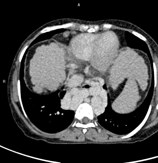
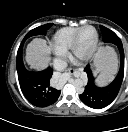
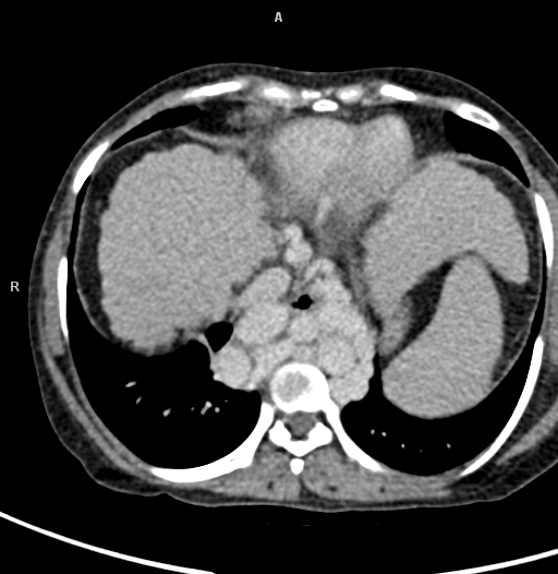
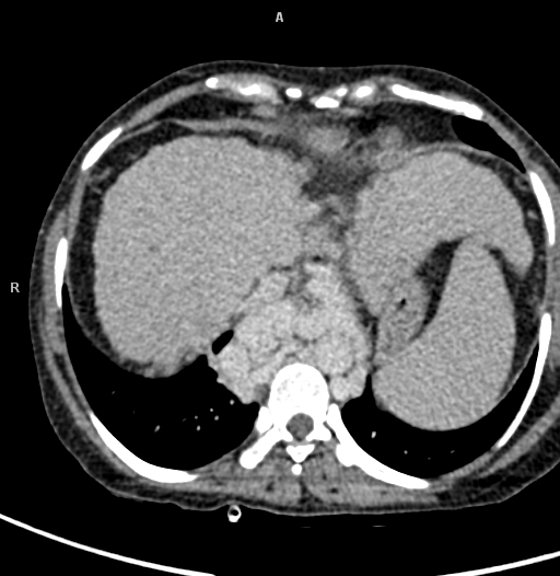
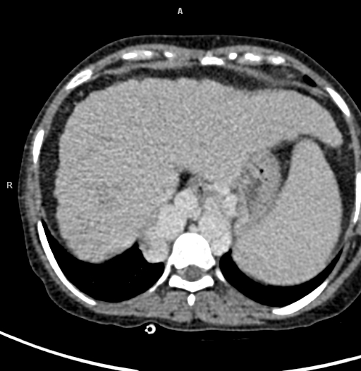
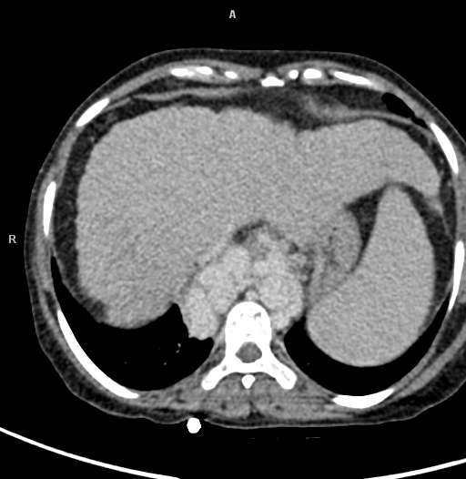
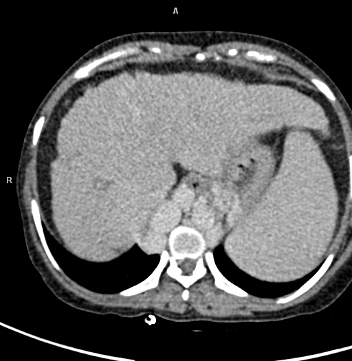
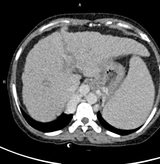
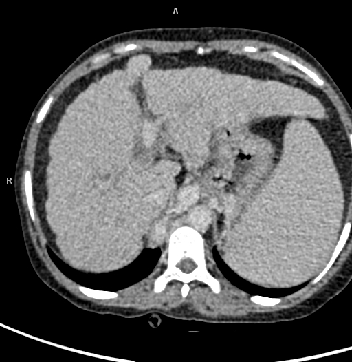
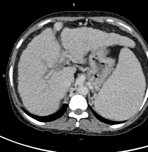
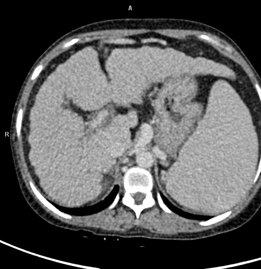
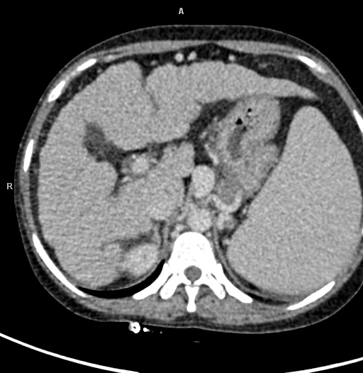
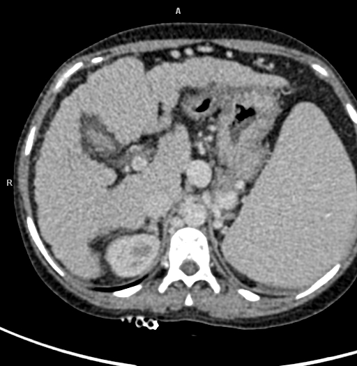
· Liver measures 11.5cm in size. Atrophy of right lobe of liver is seen with irregular and nodular surface. Heterogenous enhancement of liver parenchyma is seen.
· Portal vein appears non dilated, measures 1.2 cm at porta. Hypodense filling defect s/o thrombus is noted involving main portal vein at the portosplenic confluence extending for a length of 3.6cm causing its partial luminal occlusion.
· Splenic vein and superior mesenteric veins appear dilated and tortuous with maximum diameter measuring 2.1cm and 1.3 cm respectively.
· Multiple periportal, perigastric, peripancreatic, linorenal, splenic hilar, perisplenic anterior abdominal wall, gastroesophageal junction collaterals are noted.
· Azygus vein appears tortuous and dilated with maximum diameter measuring 1.4 cm.
· Gall bladder is minimally distended with normal wall thickness. Pericholecystic fatty infiltration is seen.
· Pancreas appears normal and shows normal contrast enhancement. CBD appears normal.
· Spleen appears enlarged, measures 19cm. It shows normal contrast enhancement. Mass effect is seen in the form of displacement of adjacent bowel loops and posteroinferior displacement of left kidney.
· Aorta, IVC, and SMA appears normal. Both adrenals are normal.
· Both kidneys appear normal in size and shows prompt nephrogram and good excretion of contrast. Both ureters appear normal in course and caliber.
· Urinary bladder appears partially distended, normal. Uterus appears normal.
· Few homogenously enhancing enlarged lymph nodes are noted in mesenteric region with largest measuring 1.2x1 cm.
· Mild free fluid is noted in abdomen and pelvis.
· Rest of visualized bowel loops appears normal.
· Visualised lung parenchyma appears normal.
· VIsualised bones appear normal.
Impression:
· Atrophy of right lobe of liver with irregular and nodular surface s/o liver parenchymal disease.
· Portal vein thrombosis with multiple portosystemic venous collaterals as described above.
· Splenomegaly.
· Mild ascitis.
Possibility of changes of portal hypertension needs to be considered.