


Pathophysiology: Multiple myeloma can cause a wide variety of problems. The proliferation of plasma cells may interfere with the normal production of blood cells, resulting in leukopenia, anemia, and thrombocytopenia. The cells may cause lytic lesions in the skeleton or in soft tissue masses. Feared complications of this malignancy are bone pain, hypercalcemia, and spinal cord compression. The aberrant antibodies that are produced lead to impaired humoral immunity, and patients have a high prevalence of infection, especially with encapsulated organisms. The overproduction of these antibodies may lead to hyperviscosity, amyloidosis, and renal failure
History: Presenting symptoms include bone pain, pathologic fractures, weakness, anemia, infection (often resulting from pneumococcal infection), hypercalcemia, spinal cord compression, or renal failure. Increasingly, physicians are identifying asymptomatic patients through routine blood screening. Typically, a large gap between the total protein and the albumin levels observed on an automated chemistry panel suggests a problem (ie, protein minus albumin equals globulin).
Histologic Findings: In patients with myeloma, plasma cells proliferate within the bone marrow, typically in sheets. Plasma cells are 2-3 times larger than typical lymphocytes; they have eccentric nuclei that are smooth (round or oval) in contour with clumped chromatin and have a perinuclear halo or pale zone. The cytoplasm is basophilic. Many descriptions of myeloma cells include characteristic, but not diagnostic, cytoplasmic inclusions, which include Mott cells, Russell bodies, grape cells, and morula cells. Bone marrow examination reveals plasma cell infiltration, often in sheets or clumps. This infiltration is different from the lymphoplasmacytic infiltration observed in patients with Waldenström macroglobulinemia.
Staging: Staging is a cumulative evaluation of all of the diagnostic information garnered and is a useful tool for stratifying the severity of patients' disease. The staging system for myeloma is somewhat complex, but it is well correlated with outcome.
Stage I involves all of the following:
Hemoglobin level greater than 10 g/d.
Calcium level less than 12 mg/dL
Radiograph showing normal bones or solitary plasmacytoma.
Low M protein values (ie, IgG <5 g/dL, IgA <3 g/dL, urine <4 g/24 h).
Stage II involves criteria that fit neither stage I nor stage III.
Stage III involves any one of the following:Hemoglobin level less than 8.5 g/dL.
Calcium level greater than 12 mg/dL.
Radiograph showing advanced lytic bone disease
High M protein value (ie, IgG >7 g/dL, IgA >5 g/dL, urine >12 g/24 h)
Subclassification A involves a creatinine level of less than 2 g/dL.
Subclassification B involves a creatinine level greater than 2 g/dL.
Stage I is associated with median survival of longer than 60 months, stage II is 41 months, and stage III is 23 months. Stage B disease has a significantly worse outcome (eg, 2-12 mo in 4 separate series).
Radiographic Findings:
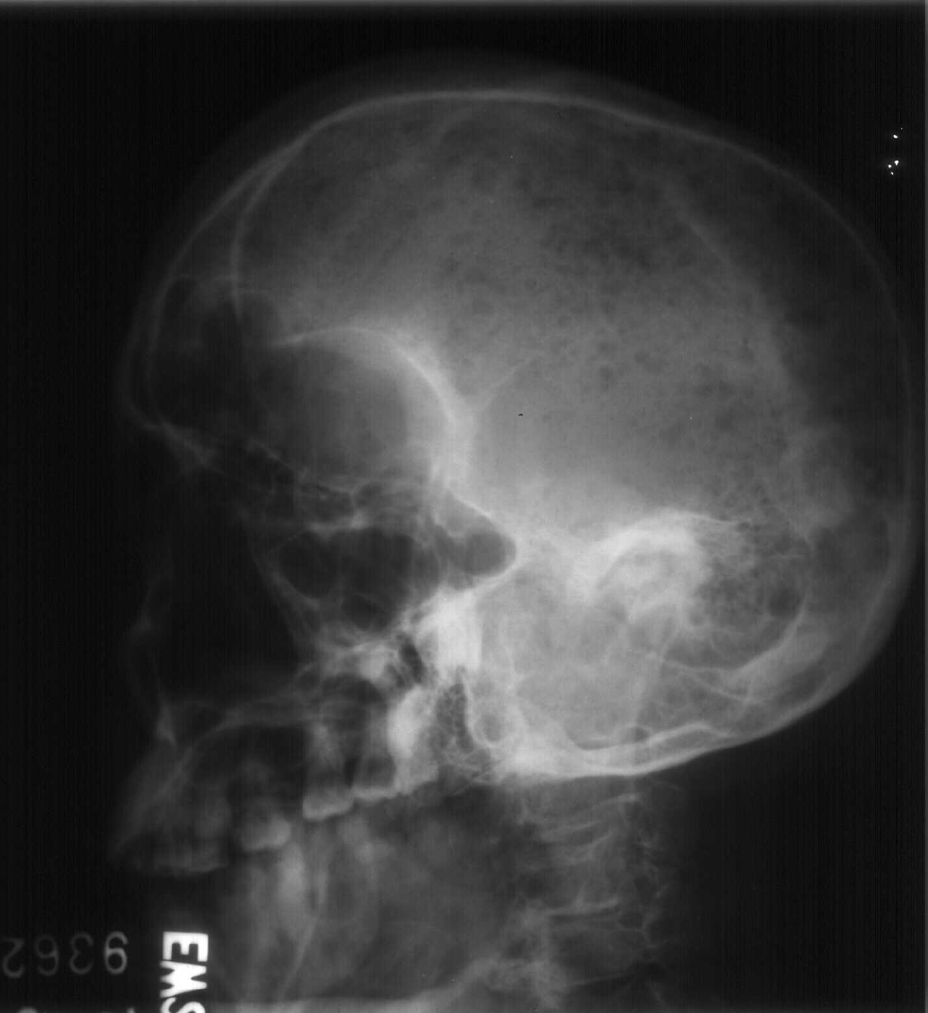
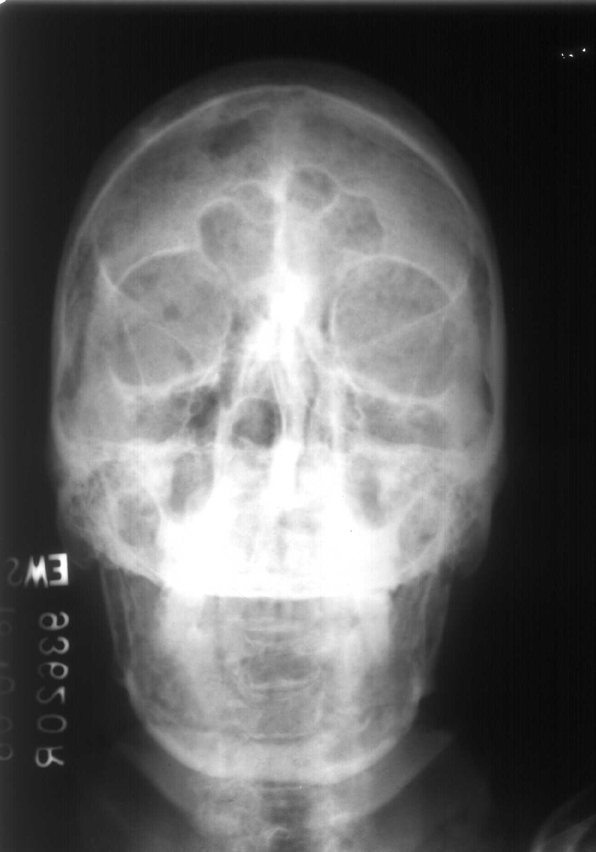
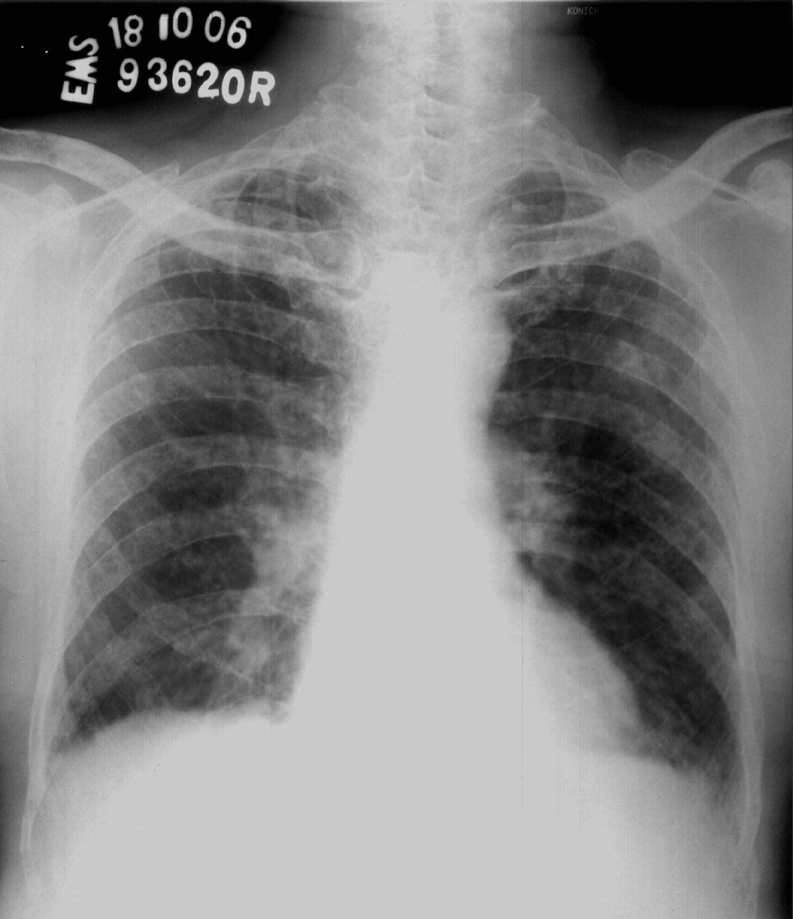
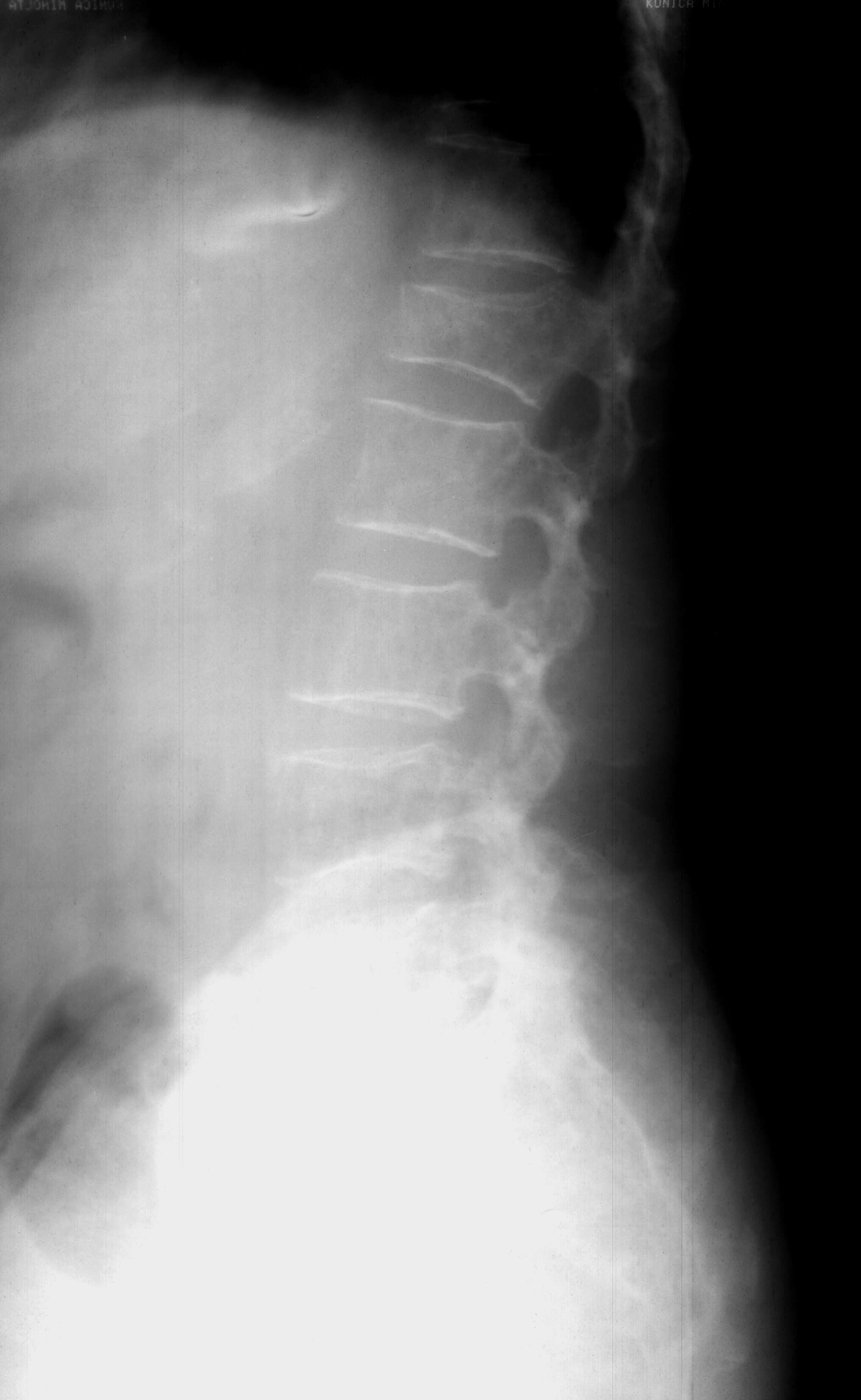
The classic radiographic appearance of multiple myeloma is that of multiple, well-circumscribed, lytic, punched-out, round lesions within the skull, spine, and pelvis.
The lesions tend to vary slightly in size. In addition, the bones of myeloma patients are, with few exceptions, diffusely demineralized. Since myeloma is a disease of the medullary compartment of the bone, more subtle lesions can be detected by the appearance of endosteal scalloping seen as slight undulation to the inner cortical margin of bone. This is suggestive of myelomatous involvement.
Although patients with advanced and extensive myeloma tend to have a number of circumscribed lytic lesions, some simply may have diffuse osteopenia on radiography.
Fewer than 10% of patients present with a single myelomatous lesion, a plasmacytoma, found on radiographs. These lesions are bubbly expansions of a single bone, often the ribs or posterior elements of the spine, and occasionally are associated with a soft-tissue mass.
A rare form of myeloma known as POEMS (polyneuropathy, organomegaly, endocrinopathy, monoclonal gammopathy, and skin changes) syndrome may demonstrate sclerotic lesions on radiographs, but this is responsible for fewer than 1% of myeloma cases. Radiographs of treated myeloma lesions also may show areas of abnormal bone architecture with sclerosis. Usually, little periosteal reaction is seen.
CT Findings:
CT depicts osseous involvement in myeloma. However the usefulness of CT has not been studied well, and CT is not required in most patients, since the standard skeletal surveys usually depict most of the lesions that CT can detect.The single clinical situation in which CT may be of value is in the patient with bone pain and a negative radiograph. In this case, demonstration of a myeloma lesion may alter therapy significantly. CT also can guide percutaneous biopsies, especially of osseous or extraosseous lesions suspected of being plasmacytomas.
MRI Findings:
MRI is potentially useful in imaging multiple myeloma because of its superior soft tissue resolution.
T1W
The typical appearance of a myeloma deposit is a round low signal intensity (relative to muscle) focus on T1-weighted images
T2W
High in signal intensity on T2-weighted sequences demonstrate the appearance of a typical myeloma lesion in the proximal humerus.
T1+C
Myeloma lesions tend to enhance somewhat with gadolinium administration. In addition, diffuse areas of replacement of the normal fatty marrow may be seen, resulting in large regions of low T1-weighted signal.
Role of Intervention: Myeloma is treated with chemotherapy and, possibly, radiation. CT may be used for percutaneous biopsy. Vertebroplasty recently has been suggested as a treatment for pathologic fractures within the spine
Staging: Staging is a cumulative evaluation of all of the diagnostic information garnered and is a useful tool for stratifying the severity of patients' disease. The staging system for myeloma is somewhat complex, but it is well correlated with outcome.
Stage I involves all of the following:
Hemoglobin level greater than 10 g/d.
Calcium level less than 12 mg/dL
Radiograph showing normal bones or solitary plasmacytoma.
Low M protein values (ie, IgG <5 g/dL, IgA <3 g/dL, urine <4 g/24 h).
Stage II involves criteria that fit neither stage I nor stage III.
Stage III involves any one of the following:Hemoglobin level less than 8.5 g/dL.
Calcium level greater than 12 mg/dL.
Radiograph showing advanced lytic bone disease
High M protein value (ie, IgG >7 g/dL, IgA >5 g/dL, urine >12 g/24 h)
Subclassification A involves a creatinine level of less than 2 g/dL.
Subclassification B involves a creatinine level greater than 2 g/dL.
Stage I is associated with median survival of longer than 60 months, stage II is 41 months, and stage III is 23 months. Stage B disease has a significantly worse outcome (eg, 2-12 mo in 4 separate series).
Radiographic Findings:
The classic radiographic appearance of multiple myeloma is that of multiple, well-circumscribed, lytic, punched-out, round lesions within the skull, spine, and pelvis.
The lesions tend to vary slightly in size. In addition, the bones of myeloma patients are, with few exceptions, diffusely demineralized. Since myeloma is a disease of the medullary compartment of the bone, more subtle lesions can be detected by the appearance of endosteal scalloping seen as slight undulation to the inner cortical margin of bone. This is suggestive of myelomatous involvement.
Although patients with advanced and extensive myeloma tend to have a number of circumscribed lytic lesions, some simply may have diffuse osteopenia on radiography.
Fewer than 10% of patients present with a single myelomatous lesion, a plasmacytoma, found on radiographs. These lesions are bubbly expansions of a single bone, often the ribs or posterior elements of the spine, and occasionally are associated with a soft-tissue mass.
A rare form of myeloma known as POEMS (polyneuropathy, organomegaly, endocrinopathy, monoclonal gammopathy, and skin changes) syndrome may demonstrate sclerotic lesions on radiographs, but this is responsible for fewer than 1% of myeloma cases. Radiographs of treated myeloma lesions also may show areas of abnormal bone architecture with sclerosis. Usually, little periosteal reaction is seen.
CT Findings:
CT depicts osseous involvement in myeloma. However the usefulness of CT has not been studied well, and CT is not required in most patients, since the standard skeletal surveys usually depict most of the lesions that CT can detect.The single clinical situation in which CT may be of value is in the patient with bone pain and a negative radiograph. In this case, demonstration of a myeloma lesion may alter therapy significantly. CT also can guide percutaneous biopsies, especially of osseous or extraosseous lesions suspected of being plasmacytomas.
MRI Findings:
MRI is potentially useful in imaging multiple myeloma because of its superior soft tissue resolution.
T1W
The typical appearance of a myeloma deposit is a round low signal intensity (relative to muscle) focus on T1-weighted images
T2W
High in signal intensity on T2-weighted sequences demonstrate the appearance of a typical myeloma lesion in the proximal humerus.
T1+C
Myeloma lesions tend to enhance somewhat with gadolinium administration. In addition, diffuse areas of replacement of the normal fatty marrow may be seen, resulting in large regions of low T1-weighted signal.
Role of Intervention: Myeloma is treated with chemotherapy and, possibly, radiation. CT may be used for percutaneous biopsy. Vertebroplasty recently has been suggested as a treatment for pathologic fractures within the spine