


The scaphoid bone is the most commonly fractured carpal bone; this injury occurs most often in young men typically those aged 15-60 years. Scaphoid fractures are rare in young children and the elderly because of the relative weakness of the distal radius compared with the scaphoid in these age groups. Scaphoid fractures are significant because a delay in diagnosis can lead to a variety of adverse outcomes that include nonunion, delayed union, decreased grip strength, decreased range of motion, and osteoarthritis of the radiocarpal joint.
Normal Anatomy:The scaphoid is a biomechanically important, boat-shaped carpal bone (from the Greek "skaphos," meaning "boat") that articulates with the distal radius, trapezium and capitate. On surface anatomy, the scaphoid is located below the anatomic snuffbox .This triangular depression is defined by the extensor and abductors of the thumb, and is easily visible when the wrist is partially ulnar deviated and the thumb abducted and extended.
The dorsal and volar branches of the radial artery provide the blood supply to the scaphoid. The primary blood supply comes from the dorsal branch of the radial artery, which divides into 2-4 branches before entering the waist of the scaphoid along the dorsal ridge. The branches course volar and proximal within the bone, supplying 70-85% of the scaphoid. The volar scaphoid branch also enters the bone as several perforators in the region of the tubercle; these supply the distal 20%-30% of the bone .
Anatomically, the scaphoid may be divided into proximal, middle (termed the waist), and distal thirds. Most of the blood supply to the scaphoid enters distally. The proximal part of the scaphoid has no blood vessels entering it and depends on vessels piercing its mid portion.
The primary mechanism of injury to the scaphoid bone is a fall on an outstretched hand. Concurrent fractures about the wrist occur in 5-12% of scaphoid fractures. The most frequently encountered fractures are radial styloid fractures, triquetrum fractures, capitate fractures, or transcarpal perilunate fracture-dislocations
Clinical Presentation: The patient complains of a deep, dull pain in the radial wrist. The pain, which often is mild, is worsened by gripping or squeezing. There may be mild wrist swelling or bruising and, possibly, fullness in the anatomic snuffbox, suggesting a wrist effusion.
Fractures can be grouped according to the anatomic location: the tubercle, the distal pole, the waist, or the proximal pole.
Scaphoid fractures can also be classified according to the plane of fracture with respect to the long axis of the scaphoid into horizontal oblique, transverse, and vertical oblique fractures .The most important classification scheme distinguishes stable scaphoid fractures and those unstable fractures .
Stable fractures are those that are incomplete, or, if they appear complete, they likely have an incompletely disrupted articular surface (the overlying cartilage is intact).
Unstable fractures are complete fractures with motion about the fracture site.
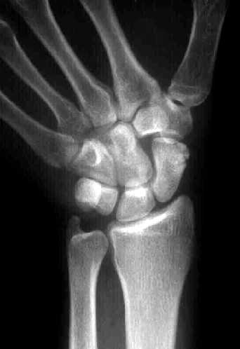
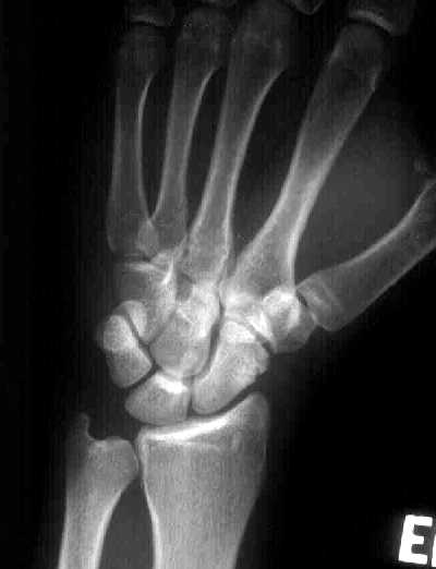
Radiographic evaluation:
The initial radiographic assessment of scaphoid fractures is performed with plain radiography using minimum of three views: PA; semipronated oblique, often with ulnar deviation; and true lateral.
To elongate the scaphoid, a scaphoid view is often obtained by positioning the wrist in ulnar deviation and angling the tube cranially by 20-40°.
A fracture typically is identified as a lucent line with at least 1 disrupted cortex.The scaphoid, or navicular, fat stripe is fat interposed between the radial collateral ligament and the tendons of the abductor pollicis longus and extensor pollicis brevis. It is visible in 90% of healthy individuals .Obliteration or displacement of the fat stripe usually occurs within 1 hour after the scaphoid fracture occurs.Because a normal fat stripe with a scaphoid fracture is exceedingly uncommon, a scaphoid fracture is virtually excluded when the scaphoid fat stripe is normal.
Angulation of the scaphoid at the fracture is often called the humpback deformity. This angulation is associated with greater likelihood of nonunion, worse clinical outcome, and arthritis.
The classic pattern of a fracture on an MRI is a linear focus of decreased signal intensity on T1-weighted images. Increased signal intensity in a distribution similar to that of the T1-weighted images is seen with T2-weighted sequences. The fracture line may be more difficult to see on T2-weighted images. Short-tau inversion recovery (STIR) or fat-suppressed T2-weighted sequences are very sensitive to edema.
Complications of scaphoid fractures may include malunion, delayed union and nonunion, and avascular necrosis (AVN). Osteonecrosis is more common in scaphoid fractures than most another bones because of the blood supply to this bone.Osteonecrosis occurs in 15-30% of all scaphoid fractures, and most of these involve the proximal pole. Its incidence increases as the fracture line becomes more proximal.
Complications of undiagnosed scaphoid fracture that leads to malunion: Chronic pain, decreased ROM, and decreased grip strength may result.
Long-standing scaphoid nonunion may result in carpal collapse. This is called a scaphoid nonunion advanced collapse (SNAC) wrist. This pattern may present 4-5 years or more than 20 years after the initial injury. Radiographic hallmarks of nonunion: sclerosis at the fracture site, a persistent lucent line that usually is wider than 2 mm, cystic cavitation, displacement of more than 1 mm, and local tenderness. The sclerotic margins, though characteristic, may become evident only after several months or years as the pseudoarthrosis matures.