


CRANIOPHARYNGIOMA
Craniopharyngiomas are benign, extra-axial, predominantly cystic, slow-growing epithelial tumors that predominately involve the sellar and suprasellar space and are presumed to arise from remnants of Rathke’s pouch epithelium. They represent 1.2% to 4.6% of all intracranial tumors but are more common in children, in whom they represent 5% to 10% of intracranial neoplasms. They can metastasize, and patients can have severe symptoms that usually require surgery.
There are 3 distinct subtypes of Craniopharyngiomas have been described. They are:-
1) Adamantinomatous tumor.
2) Papillary tumor.
3) Mixed tumor.
Craniopharyngiomas are slow growing tumors that extends superiorly and often insinuate into the third ventricle. They may also expand anteriorly under the frontal lobes (30%), posteriorly into the interpeduncular cistern and laterally into the medial aspect of the middle cranial fossa. The adamantinomatous tumors provoke an intense gliosis in the adjacent brain and become firmly adherent to adjacent brain and vessels, they have a multilobulated inner aspect with nodules of solid tissue separated by cysts of varying size. The cystic component is usually predominant in the adamantinomatous tumors and much less prominent in the papillary variety. Calcification is present in the solid portions of nearly 90% of adamantinomatous craniopharyngiomas but is much less common in papillary tumors.
On CT these tumors typically appear as heterogeneously hypodense suprasellar masses with nodular solid areas intermixed with cystic areas of varying size. More than 80% of craniopharyngiomas in children demonstrate nodular and curvilinear calcifications in the solid nodules and in the cyst walls. Papillary craniopharyngiomas in adults appear more solid and rarely demonstrate calcification. Post contrast images shows intense enhancement of the lesion.
CLINICAL FEATURES:
The most common presenting symptoms are headache, nausea, vomiting and visual disturbances. Other common findings include occulomotor palsies, bizarre scotomas, blindness, asymmetric acuity deficiencies, and optic atrophy. Hydrocephalus may result from tumor that obstructs the third ventricle. Papilledema frequently occurs. In children growth failure and headache are most common.
DIFFERENTIAL DIAGNOSIS:
1) Arachnoid cyst.
2) Astrocytoma, Brain.
3) Epidermoid, Brain.
4) Meningioma, Brain.
5) Pituitary adenoma.
6) Rathke cleft cyst.
TREATMENT:
Treatment is surgery and/or radiation therapy with intracystic chemotherapy in some pediatric patients.
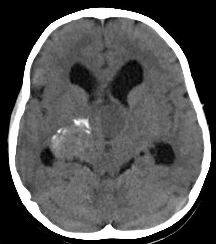
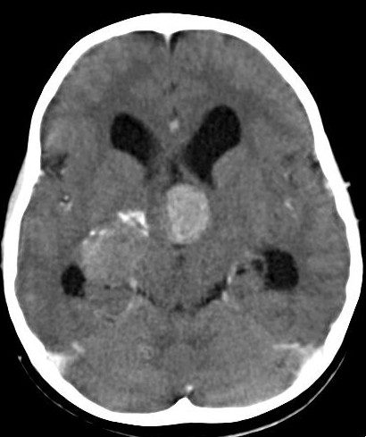
In our case CT findings are as follows:-
There is a soft tissue density mass lesion in the suprasellar region. Lesion shows isodense soft tissue component with calcification. There is nodular calcification seen along the periphery.
There is compression of the third ventricle which results in hydrocephalus.
There is also compression of the temporal horns.
Post contrast images shows intense enhancement of the lesion.
All imaging feature are suggestive of CRANIOPHARYNGIOMA.