


SPINAL MENINGIOMA
Introduction:
Meningiomas are the second most common cause of spinal tumor (in the intradural extramedullary location), second only to tumors of the nerve sheath. Meningiomas account for approximately 25% of all spinal tumors. Approximately 80% of spinal meningiomas are located in the thoracic spine, followed by cervical spine (15%), lumbar spine (3%), and the foramen magnum (2%). Most spinal meningiomas are benign. Aggressive tumors and hemangiopericytomas are rare.
Pathophysiology:
Spinal meningiomas are often located laterally or dorsolaterally in the thoracic spine. Meningiomas of the cervical and foramen magnum tend to be located ventral to the spinal cord. They arise from the arachnoid cluster cells located at the entry zone of the nerve roots or at the junction of dentate ligaments and dura mater, where the spinal arteries penetrate. For this reason, lateral tumors are more common than dorsal and ventral lesions. Most meningiomas are intradural and extramedullary. Occasionally, they can be purely extradural (7%) or intradural and extradural (6%).
Compression of the cord by the meningioma can cause deterioration of neurologic function.
Spinal meningiomas differ from intracranial meningiomas by their slightly greater proclivity for psammomatous change. In general, histopathologic features of spinal meningiomas are similar to their intracranial counterparts. Spinal meningiomas are typically globoid, and they vary in consistency depending on the extent of calcification. Multiple meningiomas are rare (2%) and most often associated with neurofibromatosis type II.
Incidence, Age and Gender:
Meningiomas are second only to nerve sheath tumors in frequency, accounting for approximately 25% of all spinal tumors. The ratio of spinal to intracranial meningiomas is about 1:8. The peak incidence is in the fifth and sixth decades. More than 80% of spinal meningiomas occur in women. Multiple spinal meningiomas are rare.
Location:
Spinal meningiomas often are located laterally or dorsolaterally in the spinal canal. 90% of spinal meningiomas are intradural, whereas 5% are “dumbshell†or extradural lesions. They are believed to arise from the arachnoid cluster cells, and therefore, they are located at the entry zone of the nerve roots or the junction of the dentate ligaments and dura mater. The spinal cord is typically compressed and displaced away from the lesion. The subarachnoid space above and below the mass lesion is widened, with cerebrospinal fluid capping the lesion from above and below. On occasion, they can be purely extradural (7%) or intradural and extradural (6%).
Clinical presentation:
The clinical course may be insidious, and symptoms are often confused with symptoms of other lesions of the spine, peripheral nervous system, and thorax.
Symptoms produced by meningiomas are secondary to their broad dural attachment and the gradual growth of the tumor with compression of the cord.
Symptoms are:
1) Motor and sensory deficits. ( 90% and 60% of patients )
2) Sphincter dysfunction and pain. ( local, radicular, funicular, occur in about 50% of cases )
3) A high incidence of Brown-Sequard syndrome is seen, with ipsilateral paralysis, decreased tactile and deep sensation, and a contralateral deficit in pain and temperature sensation.
4) Patients most frequently complain of regional back pain, especially at night, followed by sensorimotor changes and, eventually, bowel and bladder dysfunction.
IMAGING FEATURES:
CT Findings:
NECT scans may disclose an extradural or “dumbshell “mass that is iso- or moderately hyperdense compared to muscle.
CT scans obtained with the intravenous injection of contrast material may show a homogeneous enhancing tumor.
Myelography or CT myelography is required to demonstrate the intradural extramedullary location of the mass.
The spinal cord is displaced away from the lesion and usually compressed. A sharp meniscus is seen where the contrast agent caps the lesion from above and below. The subarachnoid space on the side of the lesion is widened.
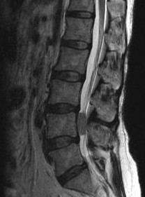
MR Findings:
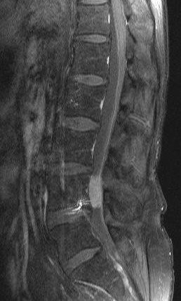
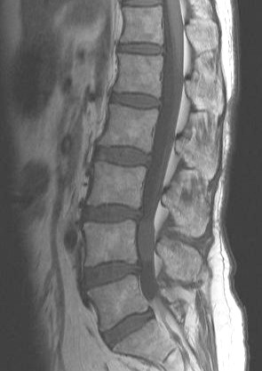
MRI demonstrates the intradural extramedullary location of meningiomas. Lesions are usually isointense to spinal cord on both T1-weighted and T2-weighted images. Lesions are sometimes hypointense on T1-weighted images and hyperintense on T2-weighted images. Homogeneous intense enhancement of the lesion is seen after an intravenous injection of gadolinium-based contrast agent. Occasionally, densely calcified meningiomas are profoundly hypointense on MR and show only minimal contrast enhancement.
Most spinal meningiomas demonstrate broad-based dural attachment. On occasion, a densely calcified meningioma may demonstrate hypointensity on both T1-weighted and T2-weighted images. The spinal cord is displaced away from the lesion and usually compressed. The subarachnoid space above and below the lesion is widened, and a meniscus capping the lesion may be seen.
MRI is more reliable than CT in demonstrating Spinal meningioma
Ultrasound Findings:
Intraoperative ultrasonography may be performed after laminectomy as a tool to localize the mass, which is usually echogenic.
Angiography Findings:
Spinal angiography usually shows a hypervascular lesion with tumor blush. More important, spinal angiography is performed to identify the location of the artery of Adamkiewicz, which is usually in the lower thoracic region, most frequently on the left side. Care must be taken not to damage the artery during surgery because the artery supplies the spinal cord. Spinal angiography is performed for surgical planning if the meningioma is in the lower thoracic region, regardless of the side of the meningioma.
Treatment:
Surgical resection of the tumor.
Intervention: In highly vascular spinal meningiomas, preoperative embolization with particles may be performed to minimize blood loss during surgery. However, this procedure is usually not necessary because highly vascular spinal meningiomas are uncommon.
Differential Diagnosis:
1) Astrocytoma
2) Dermoid tumor
3) Ependymoma
Other problems to be considered.
Schwannoma
Intradural metastasis
Teratoma
Exophytic astrocytoma
Exophytic ependymoma
Intradural metastasis
Teratoma
Exophytic astrocytoma
Exophytic ependymoma