


Rickets
Rickets is a disease caused by deficiency of Vitamin D leading to bony deformities and hypocalcaemia.
Rickets is a disease caused by deficiency of Vitamin D leading to bony deformities and hypocalcaemia.
Pathophysiological basis of radiographic findings in Rickets
Loss of orderly maturation and mineralization of cartilage cells at the growth plate resulting from Vitamin D deficiency is ‘Rickets’. Thus, Rickets is like osteomalacia in a growing skeleton.
Inherent to all rachitic syndromes are osteomalacic changes in portions of skeleton that contains mineralized bone (Mature bone-both compact & spongy).
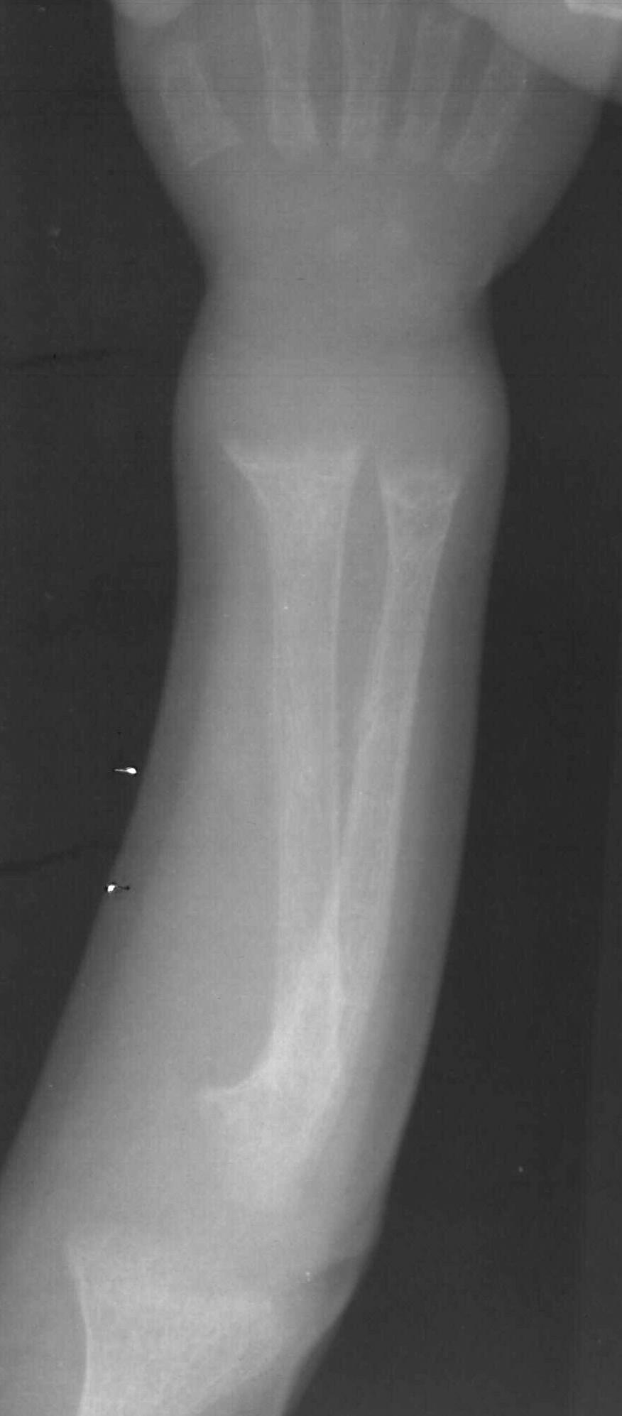
The skeletal effects are due to lack of calcification of osteoid. The most obvious change are at ‘metaphysis’- where rapid growth is occurring.
First change to appear is a ‘loss of normal zone of provisional calcification’ adjacent to metaphysis. This begins as an indistinctness of the metaphyseal margin, progressing to a ‘frayed’ appearance with widening of the growth plate, due to lack of calcification of metaphyseal bone. Weight bearing & stress on uncalcified bone gives rise to ‘splaying’ & ‘cupping’ of metaphysis. A similar but less marked effect occurs in subperiosteal layer , which may cause lack of distinctness of cortical margin. Eventually a generalized reduction in bone density is seen. In the epiphysis – there may be some haziness of cortical margin. Thus all findings in Rickets occur due to failure of calcification & abnormal demineralization
Healing Rickets
The first changes of rickets appear in rapidly growing distal ends of ulna & radius (wrist & knee are commonly involved due to more use). Rarefaction of provisional zone of calcification with widening of epiphysis- diaphysis distance is first to appear. Following treatment there is ossification of provisional zone of calcification.
However, early signs of Rickets should be always looked for e.g. On chest X-ray, humoral head may show certain early radiological changes
Skull & spine changes in Rickets
Skull changes | -Pronounced calvarial demineralization
(Even facial bones are involved)
| -Basilar Invagination
| -Indistinct sutural margin
| -Delayed tooth eruption
| -Premature craniostenosis
| -Craniotabes
| -Calvarial thickening following treatment
Spine changes | -Scoliosis
| -Biconcave vertebral bodies
| -Triradiatic pelvis.
(Even facial bones are involved)
| -Basilar Invagination
| -Indistinct sutural margin
| -Delayed tooth eruption
| -Premature craniostenosis
| -Craniotabes
| -Calvarial thickening following treatment
Spine changes | -Scoliosis
| -Biconcave vertebral bodies
| -Triradiatic pelvis.
Imaging signs of healing Rickets
With treatment, there is regression of radiological findings seen in Rickets. During a stage of healing, there is extensive periosteal new bone formation as a reflection of ossification occurring along the cornices of diaphyses. Also seen during healing are radiolucent bands at metaphyses of long bones & cupping of metaphyses becomes clearly prominent. As healing progresses remodeling of bowing deformities occur & in skull characteristic bossing of frontal & parietal bones becomes apparent with premature closure of sutures.
With Renal Tubular acidosis, a peculiar brush border along the metaphysis is seen in the healing phase.
Vitamin D Resistant Rickets on imaging
Vitamin D Resistant Rickets is found in older children (>30months of age). Patient is short, stocky & bow legged. Ectopic calcifications & ossification in axial/appendicular skeleton along with occasional sclerotic changes are among the identifying radiographic features. Bowing of legs & shortening of long bones are more pronounced. Bones are more sclerotic.
Neonatal Rickets
In first 2 years of life, incidence of Rickets is 5 to 20%. Neonatal rickets is believed to be of multifactorial origin. Major contributing factors are related to nutrition, immaturity of enzyme systems & iatrogenic / metabolic factors. Premature infants of low birth weight are primarily affected. Bony changes appear around 2 months of age. The most frequent causes of Rickets in patients under 6 months of age also include conditions like Biliary Atresia.
In first 2 years of life, incidence of Rickets is 5 to 20%. Neonatal rickets is believed to be of multifactorial origin. Major contributing factors are related to nutrition, immaturity of enzyme systems & iatrogenic / metabolic factors. Premature infants of low birth weight are primarily affected. Bony changes appear around 2 months of age. The most frequent causes of Rickets in patients under 6 months of age also include conditions like Biliary Atresia.
Sequelae post-rickets
Complete healing & restoration of normal structure are rule in rickets, however distortion/ sclerosis of spongiosa in affected segment may occur after healing & may remain visible for several years. Cortical thickening of segments of bone involved during active stage also may persist. Angulation deformities secondary to pathological fractures result in deformities like knock-knee, bow leg and saber shin.