


Renal Oncocytoma.
Oncocytoma is the most common benign solid renal tumor. Oncocytomas originate from the intercalated cells of the renal collecting duct.
Anatomy
The kidneys are retroperitoneal organs that are enclosed in a fibrous capsule and surrounded by perirenal fat. The anterior pararenal fascia separates the kidneys from the pancreas, and the posterior pararenal fascia demarcates the paraspinal muscles from the kidneys. The adrenal glands are located superomedially.
Each kidney is supplied by a main renal artery that arises directly from the aorta. Multiple renal arteries are common, and these play an important role in nephron-sparing surgeries (partial nephrectomy). A single renal vein drains each kidney, and multiple renal veins are uncommon. Lymphatics from the kidney drain into the lymph nodes in the renal hilum, which in turn drain into nodes in the para-aortic region.
Clinical Details
Approximately 56-91% of the oncocytomas are incidentally detected on imaging studies that have been performed for another indication. However, 17-21% of affected patients present with symptoms such as hematuria, flank pain, and an abdominal mass. In patients who present with symptoms, hematuria is more common than mass-like findings.
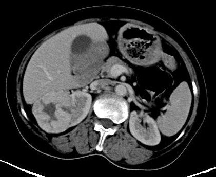
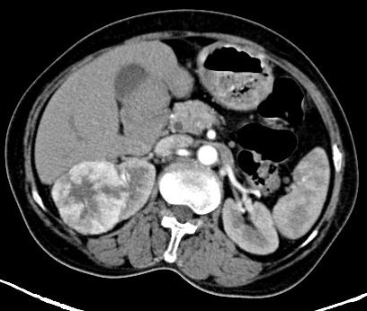
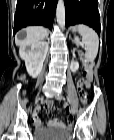
CT Findings -
On nonenhanced CT scans, oncocytomas appear isoattenuating or slightly hyperattenuating relative to the kidney parenchyma. On contrast-enhanced CT scans that are obtained during the nephrographic phase, the mass appears less attenuating than the renal parenchyma.
On nonenhanced CT scans, oncocytomas appear isoattenuating or slightly hyperattenuating relative to the kidney parenchyma. On contrast-enhanced CT scans that are obtained during the nephrographic phase, the mass appears less attenuating than the renal parenchyma.
Oncocytomas are well encapsulated and have distinct margins, a smooth contour, and a homogeneous appearance. The tumors may range in size from 3 to 10 cm, and in symptomatic patients, the lesions are most often larger than 5 cm.
A central hypoattenuating scar may be observed in 33% of cases, but this scar cannot be differentiated from the central necrosis commonly found in RCC. With the advent of multisection CT scanning, high-resolution thin sections through the kidneys may improve detection of the central scar.
Calcification, necrosis, and hemorrhage are rare with oncocytomas. Typically, features of a malignant tumor—such as invasion or infiltration into the perinephric fat, collecting system, or vessels—are absent. Likewise, regional lymphadenopathy and metastases are not encountered in patients with oncocytoma. Occasionally, multifocal or bilateral tumors may be found.
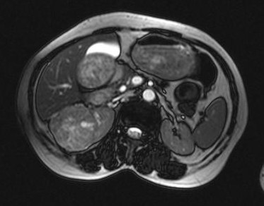
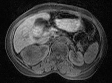
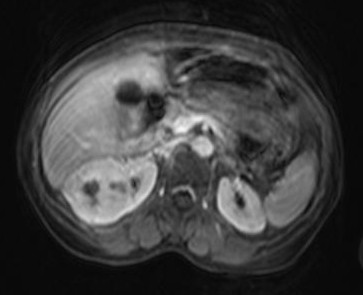
MR Findings -
On nonenhanced T1-weighted MRIs, oncocytomas are well-defined, homogeneous masses. They may appear isointense to hypointense relative to the renal cortex. On T2-weighted images, the tumors are typically isointense to slightly hypointense; however, slight T2 hyperintensity has also been reported.
When present, the tumor's scar may be seen as a hypointense, stellate area in the center of the lesion on T1- and T2-weighted MRIs. However, tumor necrosis, a common feature of malignant masses, appears hypointense on T1-weighted images and hyperintense on T2-weighted images. Rarely, the central scar may appear bright on T2-weighted images.
After the intravenous administration of gadopentetate dimeglumine contrast material, oncocytomas show homogeneous enhancement, with a nonenhancing central scar.
Angiography Findings -
With the advent of CT scanning, routine angiography is not performed to diagnose renal masses. However, the classic angiographic findings for oncocytomas include a spoke-wheel arrangement of tumoral vessels, homogeneous tumoral contrast during the capillary phase, sharp demarcation from the kidney and surrounding areas, and a peritumoral halo (lucent-rim sign). Bizarre neoplastic vessels are conspicuously absent, which is in contrast to RCC.