


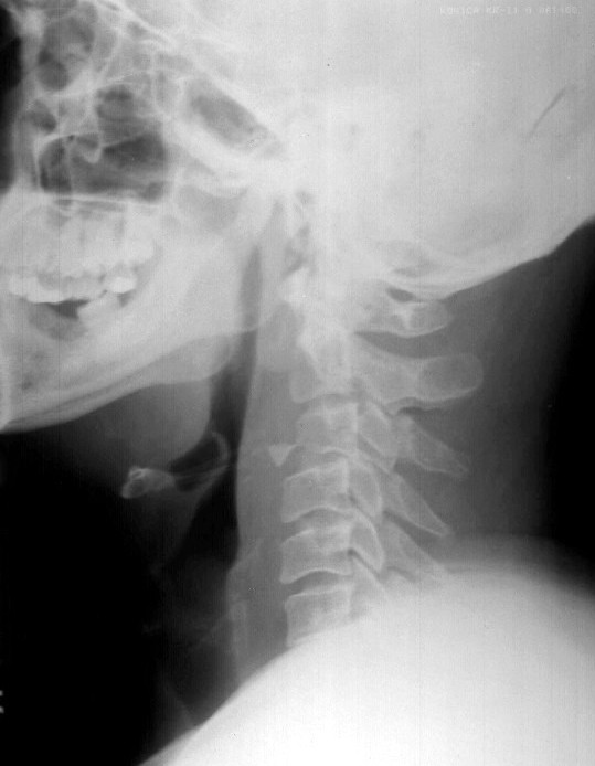
Flexion teardrop fracture
A flexion teardrop fracture occurs when flexion of the spine, along with vertical axial compression, causes a fracture of the anteroinferior aspect of the vertebral body. This fragment is displaced anteriorly and resembles a teardrop. For this fragment to be produced significant posterior ligamentous disruption must occur. Since the fragment displaces anteriorly, a significant degree of anterior ligamentous disruption exists.
This injury involves disruption of all 3 columns, making this an extremely unstable fracture that frequently is associated with spinal cord injury. Initial management is application of traction with cervical tongs.
Extension teardrop fracture
As with flexion teardrop fracture, extension teardrop fracture also manifests with a displaced anteroinferior bony fragment. This fracture occurs when the anterior longitudinal ligament pulls fragment away from the inferior aspect of the vertebra because of sudden hyperextension. The fragment is a true avulsion, in contrast to the flexion teardrop fracture in which the fragment is produced by compression of the anterior vertebral aspect due to hyperflexion.
The fracture is common after diving accidents and tends to occur at lower cervical levels. It also may be associated with the central cord syndrome due to buckling of the ligamenta flava into spinal canal during the hyperextension phase of injury.
This injury is stable in flexion but highly unstable in extension. Initial management is avoidance of iatrogenic extension and cervical traction with tongs.
Imaging Studies:
- Radiographic evaluation
- A standard trauma series is composed of 5 views: cross-table lateral, swimmer's, oblique, odontoid, and anteroposterior.
- Cross-table lateral view
- Approximately 85-90% of cervical spine injuries are evident in lateral view, making it the most useful view from a clinical standpoint.
- A technically acceptable lateral view shows all 7 vertebral bodies and the cervicothoracic junction. Approach analysis of this view methodically to avoid missing significant pathology.
- Check alignment of cervical spine by following 3 imaginary contour lines.
- The first line connects the anterior margins of all the vertebrae and is referred to as the anterior contour line.
- The second line should connect the posterior aspect of all vertebrae in a similar way and is referred to as the posterior contour line.
- The third line should connect the bases of the spinous processes and is referred to as the spinolaminar contour line.
- Each of these lines should form a smooth lordotic curve. Suspect bony or ligamentous injury if disruption is seen in the contour lines.
- An exception occurs in young children who, because of immature muscular development, may have a benign pseudosubluxation in the upper cervical spine. An imaginary straight line should connect the points bisecting the base of the spinous processes of C1, C2, and C3. In pseudosubluxation, these imaginary points should not be displaced more than 2 mm in front of or behind the straight line.
- Check individual vertebrae thoroughly for obvious fracture or changes in bone density. Areas of decreased bone density are seen in patients with osteoporosis, osteomalacia, or osteolytic lesions and may represent weak areas predisposed to injury. Areas of increased bony density may be seen with osteoblastic lesions or may represent compression fractures of an acute nature.
- Look for soft tissue changes in predental and prevertebral spaces. The predental space, also known as the atlantodental interval, is the distance between the anterior aspect of the odontoid and the posterior aspect of the anterior arch of C1 . This space should be no more than 3 mm in an adult and 5 mm in a child. Suspect transverse ligament disruption if these limits are exceeded.
- Prevertebral space extends between the anterior border of the vertebra to the posterior wall of the pharynx in the upper vertebral level (C2-C4) or to the trachea in the lower vertebral level (C6).
- At the level of C2, prevertebral space should not exceed 7 mm.
- At the level of C3 and C4, it should not exceed 5 mm, or it should be less than half the width of the involved vertebrae.
- At the level of C6, prevertebral space is widened by the presence of the esophagus and cricopharyngeal muscle. At this level, the space should be no more than 22 mm in adults or 14 mm in children younger than 15 years.
- Children younger than 24 months may exhibit a physiologic widening of the prevertebral space during expiration; therefore, obtain images in small children during inspiration to assess prevertebral space adequately.
- If the prevertebral space is widened at any level, a hematoma secondary to a fracture is the most likely diagnosis.
- Check for fanning of the spinous processes. This is evident as an exaggerated widening of the space between 2 spinous process tips and suggests posterior ligamentous disruption.
- Check for an abrupt change in angulation of greater than 11 degrees at a single interspace. This also suggests bony injury with possible ligamentous involvement.
- Swimmer's view
- Occasionally, it is impossible to fully visualize all 7 cervical vertebrae and, more importantly, the cervicothoracic junction in a true lateral image.
- Failure to fully visualize these areas has resulted in patient morbidity and successful malpractice litigation against emergency physicians.
- A swimmer's or transaxillary view adequately exposes these areas for scrutiny.
- Oblique view
- This view also is considered a laminar view because most pathologic conditions assessed on it manifest with some disruption in the normal overlapping appearance of the vertebral laminae.
- The normal structural appearance of the laminae is described as shingles on a roof, forming a regular elliptical curve with equal interlaminar spaces.
- If interlaminar space between 2 continuous laminae is increased, suspect subluxation of the involved vertebrae.
- Similarly, if the expected tiling of shingles is disrupted, suspect a unilateral facet dislocation.
- A posterior laminar fracture should be evident as disruption of the body of a single shingle.
- Odontoid view
- This view is used to evaluate an area that is difficult to visualize in the cross-table lateral view because of shadow superimposition.
- The most important structural relationship to evaluate in this view is alignment of the lateral masses of C1 with respect to the odontoid process.
- Masses should be bilaterally symmetric with the dens and odontoid process and must be checked for fractures or lateral displacement.
- Assess symmetry of the interspace between C1 and C2.
- Anteroposterior view
- This is the least useful view from a clinical standpoint.
- A straight line should connect the spinous processes bisecting the cervical spine. If this is not seen, consider a rotation injury (ie, unilateral facet dislocation). Also consider a clay shoveler fracture if a spinous process appears vertically split.