


Renal injuries occur in 15 to 40% of patients with abdominal trauma.
In patients with multivisceral trauma requiring emergent laparotomy, imaging evaluation is usually limited to emergency excretory urography prior to surgery. In more stable patients, CT allows accurate diagnosis and staging of major renal injuries.
Advantages of CT in renal trauma
CT can determine:
Ø The depth of cortical laceration
Ø The amount of infarcted renal parenchyma
Ø The extent of perirenal hemorrhage
Ø The status of the renal collecting system and the vascular pedicle.
Although excretory urography is the most cost-effective screening modality in the evaluation of stable patients with isolated flank trauma, its accuracy falls significantly with more severe renal injuries. The majority of patients with extensive renal injuries are not adequately staged by excretory urography alone. Therefore, contrast-enhanced CT should be performed in patients with suspected major renal trauma, multivisceral injuries or inadequate staging with excretory urography.
CT Based Grading of renal trauma:
GRADE I:
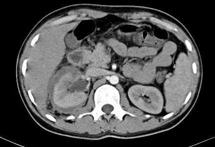
o Intrarenal hematoma or contusion
· Ill defined, round or ovoid lesion
· Parenchymal phase: Decrease enhancement relative to normal kidney
· Delayed phase: Hyperdense due to urine stasis and clot filled tubules
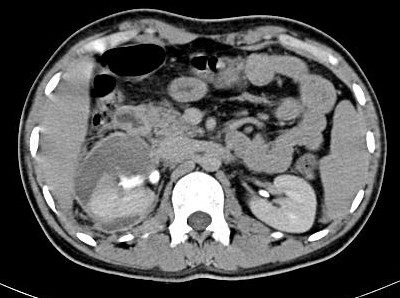
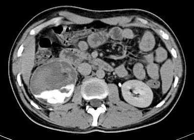
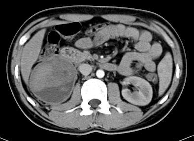
o Subcapsular hematoma
· Round or elliptic fluid collection (40-70 HU clotted blood )
o Minor lacerations: Small linear hypodense areas in periphery
o Limited perinephric hematoma: Adjacent to laceration
o Subsegmental cortical infarct
GRADE II:
o Major laceration through cortex extending to medulla
o When laceration extends into the collecting system
· Nephrographic phase: Large, distracted renal fracture
· Excretory phase: Contrast extravasation into perinephric space
· Antegrade filling of ureter (+/-)
o Segmental renal infarct: Sharpely demarcated wedge shaped area of decreased enhancement.
GRADE III:
o Multiple renal lacerations and vascular injury
· Nephrogenic phase: Several irregular, linear or band like interpolar hypodense areas with or without areas of active arterial contrast extravasation
o Subacute infarction
· “Cortical rim†sign: Preserved capsular or subcapsular enhancement
· Seen 6-8 hrs after infarction
o “Shattered infarctionâ€
· Segmental infarction: Non-enhancing wedge-shaped area
· Global infarction (nonenhancement) + no perinephric hematoma ( RA thrombosis )
· Global infarction (nonenhancement) + perinephric hematoma ( RA avulsion )
· Ureter and PUJ are patent
GRADE IV:
o Ureteropelvic junction: Complete transaction (avulsion) or laceration
o Circumferential urinoma around the affected kidney