


PIGMENTED VILLONODULAR SYNOVITIS
Jaffe, Lichtenstein and Sutro first described the disease process they termed pigmented villonodular synovitis in 1941.
Pigmented villonodular synovitis (PVNS) is a benign proliferative disorder of uncertain etiology that affects synovial lined joints, bursae, and tendon sheaths. The disorder results in various degrees of villous and/or nodular changes in the affected structures
The disease usually is monoarticular. The joint most commonly affected is the knee followed by the hips, ankle, shoulder and elbow although any synovial joint may be affected.
There are several types of pigmented villonodular synovitis:
A) Localized pedunculated villonodular synovitis (LPVNS).This type typically involves the knee causing a locking or clicking sensation in the joint. Detection and diagnosis are clinically difficult . Magnetic Resonance (MRI) is a valuable tool for the diagnosis.
B) Diffuse pigmented villonodular synovitis. (DPVNS)Tends to affect the knee but may also affect the hip or ankle. This type can mimic rheumatoid arthritis.
When the lesion arises from a tendon sheath, it is usually localized and discrete and may be termed giant cell tumour of tendon sheath (GCTTS) . GCTTS differs in location as it is classically extra-articular, located around the tendon sheaths of the hands and usually measures less than 2 cm in size
Findings on plain radiographs are not specific.
- In the diffuse form, PVNS presents as painless, monoarticular joint swelling.
- Findings of mineralization of the bones around the lesion are normal until late in the disease, with preservation of the cartilage space and no calcifications.
- Well-corticated pressure erosions (saucerization) and cysts may occur on either side or both sides of the joint.
- Secondary degenerative changes may occur in the affected joint in long-standing disease, with concentric cartilage space narrowing, subchondral cyst, and osteophyte formation.
- The diffuse form presents with joint effusion/soft-tissue swelling. Occasionally, the effusion is dense due to the presence of hemosiderin.
- The nodular form most commonly results in localized swelling of the palmar aspect of a finger.
CT scan findings are as follows:
- Secondary to the presence of intracellular and extracellular hemosiderin, lesions have high attenuation and appear hyperdense on CT scans.
- Because of improved tissue contrast, CT scanning is valuable in delineating bone cysts and erosions.
- Affected synovium is hypervascular and generally enhances following administration of radiographic contrast.
- CT scanning is useful for needle biopsy guidance when a histologic diagnosis is required.
- CT scanning is especially useful for preoperative planning.
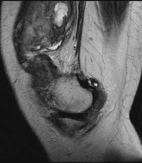
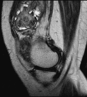
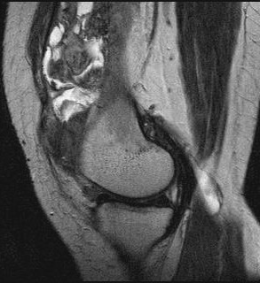
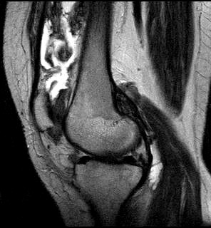
MRI findings are as follows:
- Characteristic findings include nodular intra-articular masses that demonstrate low signal intensity on T1-, T2-, and proton density–weighted sequences. Low signal intensity is due to hemosiderin deposits within the affected tissue and is accentuated on T2-weighted sequences, most notably on gradient-recalled echo sequences.
- The presence of fat signal also is apparent secondary to the presence of lipid-laden macrophages, resulting in focal regions of high signal intensity on T1-weighted images and intermediate signal on T2-weighted images.
- Hyperplastic and hypervascular synovium enhances following intravascular administration of gadolinium chelates.