


Proatlantal intersegmental artery (PIA] Type 1
Persistent caroticovertebral embryonic anastomosis was first named by Padget the "persistent primitive proatlantal intersegmental artery." Later, Lasjaunias et al called it the "proatlantal artery I." This artery is extremely rare .
The proatlantal intersegmental artery (PIA) is one of the primitive carotid-basilar anastomoses that exist transiently in the fetus. The PIA (also known as suboccipital artery) originates from the cervical internal carotid artery (ICA, type I) or external carotid artery (ECA, type II), runs upward, then curves dorsally to rest on the superior aspect of the transverse process of the atlas. It usually joins the V3 segment of vertebral artery (VA) or continues as the VA before entering the foramen magnum. Persistence of this artery in adulthood is rare and often an incidental finding. However, cerebral aneurysm, atypical cerebral infarct, intracranial hemorrhage, syncope, and headache have been attributed to the occasional persistent PIA in adults
Embryology
At the 4–5-mm embryonic stage, the hindbrain is supplied by two parallel neural arteries. These two neural arteries supply blood from the carotid system via trigeminal, otic, hypoglossal, and proatlantal arteries As the posterior communicating arteries develop, three of the four anastomoses—otic, hypoglossal, and trigeminal arteries—regress . The life span of these arteries is about a week. The proatlantal arteries persist until the vertebral arteries develop.
During the 7 to 12 mm embryonic stage, the vertebral arteries are formed from transverse anastomoses between adjacent cervical intersegmental arteries, beginning with the proatlantal intersegmental artery and proceeding downward to the C6 intersegmental artery, which forms the origin of adult vertebral artery and subclavian artery Part of the proatlantal artery becomes the horizontal portion of the vertebral artery . The horizontal and distal portions of occipital artery are also derived from the proatlantal artery
Anatomy
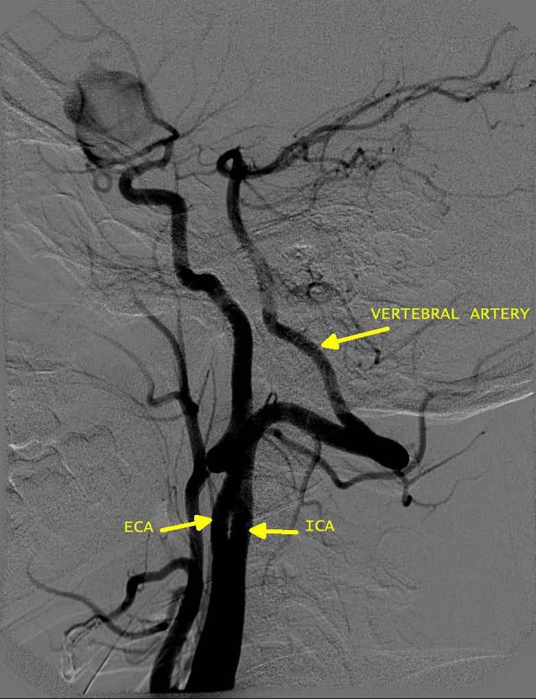
A. Persistent proatlantal artery type I (PPA 1) arises from the caudal part of the internal carotid artery and courses along the anterior aspect of the vertebral bodies to the level of the occipitoatlantal space before coursing dorsally. The vessel extends to the posterior aspect of atlas with a horizontal sweep characteristic of a type 1 proatlantal artery before turning upward to join the horizontal segment of the vertebral artery.
Anatomy
A. Persistent proatlantal artery type I (PPA 1) arises from the caudal part of the internal carotid artery and courses along the anterior aspect of the vertebral bodies to the level of the occipitoatlantal space before coursing dorsally. The vessel extends to the posterior aspect of atlas with a horizontal sweep characteristic of a type 1 proatlantal artery before turning upward to join the horizontal segment of the vertebral artery.
B.Persistent proatlantal artery type II (PPA 2) arises from the external carotid artery; it crosses the C1 or C2 vertebra obliquely. Both PPA-1 and PPA-2 enter the skull via the foramen magnum
DIFFERENTIAL
The type I proatlantal artery should be differentiated from a more frequently seen persistent hypoglossal artery. Many criteria have been established to differentiate the two arteries.
(1) The hypoglossal artery usually rises from the internal carotid artery at the level of the Cl or C1/2 interspace. The proatlantal artery I rises at the level of the C2 or C3 interspace. Thus, the hypoglossal artery leaves the internal carotid artery at a higher level than the proatlantal artery I.
(2) The proatlantal artery I curves sharply dorsally to course in the occipitoatlantal space. The suboccipital horizontal course of the proatlantal artery I and the vertebral artery is identical. However, the proatlantal artery I does not pass through the transverse process of any vertebra.
(3)The hypoglossal artery has a more vertical course than the proatlantal artery I. It lacks the suboccipital horizontal sweep characteristic of the vertebral and proatlantal arteries and never extends as far posteriorly as the proatlantal artery.
(4) The proatlantal artery enters the skull through the foramen magnum, as does the vertebral artery. The hypoglossal artery enters the skull through the hypoglossal canal. While it passes through the hypoglossal canal, it displays a small dorsal curve. This small curve must not be confused with the horizontal suboccipital course characteristic of the proatlantal artery I, which is related to the transverse process of Cl.
OTHER PERSISTENT ARTERIES
(A) Persistent hypoglossal artery - The persistent hypoglossal artery connects the distal cervical ICA to the proximal basilar artery via the hypoglossal canal. The persistent primitive hypoglossal artery is a form of segmental artery anomaly characterized by a dorsoventral anastomosis during early foetal life. . It was first described in 1889 by Batujeff incidentally during an autopsy. In 1922 Oertel termed it the “hypoglossal artery†because this artery passes through the hypoglossal canal together with the hypoglossal nerve.Later, in 1950, Lindgreen described PPHA by angiography .
The diagnosis and definition of PPHA is grounded on four criteria, according to specific angiographic findings first proposed by Lie and later revised by Brismar:
1: the PPHA leaves the internal carotid artery as a large extracranial branch at the level of C1 to C3 .
2: the PPHA enters the skull through the hypoglossal canal.
3: the basilar artery is filled only beyond the point where the artery joins it.
4: the posterior communicating arteries are absent or the ipsilateral vertebral artery may be hypoplastic or aplastic.
(B) Persistent trigeminal artery - Persistent Trigeminal Artery is a developmental anomaly in which the short, wide fetal connection between the cavernous carotid and upper third of the basilar artery does not regress. It is located below the posterior communicating artery
TYPES
In the Saltzman type I, the trigeminal artery supplies the entire vertebrobasilar system distal to the anastomosis. The basilar artery proximal to the anastomosis is usually hypoplastic, and the posterior communicating artery is often absent.
In the Saltzman type II, the anastomosis mainly supplies the superior cerebellar arteries on both sides, and the ipsilateral posterior cerebral artery is supplied through a patent posterior communicating artery.
(C)Persistent otic artery - The otic connects the cervical ICA to the vertebral artery in the region of the CN12. It may compress CN7 and 8
(C)Persistent otic artery - The otic connects the cervical ICA to the vertebral artery in the region of the CN12. It may compress CN7 and 8