


Ameloblastoma
It is an entirely epithelial tumor arising from the dental lamina, Hertwig sheath, the enamel organ, or the lining of dental follicles.
Location
Mandible: Usually centered in 3rd molar, mandibular ramus region.
Maxilla: Usually centered in premolar - 1st molar region.
Affects maxillary sinus before nose.
Most commonly presents in 30-50 year olds. The lesion is distributed equally between males and females. Although ameloblastoma generally is not classified as a malignant lesion (a rare malignant variant exists), it is extremely aggressive and infiltrative.
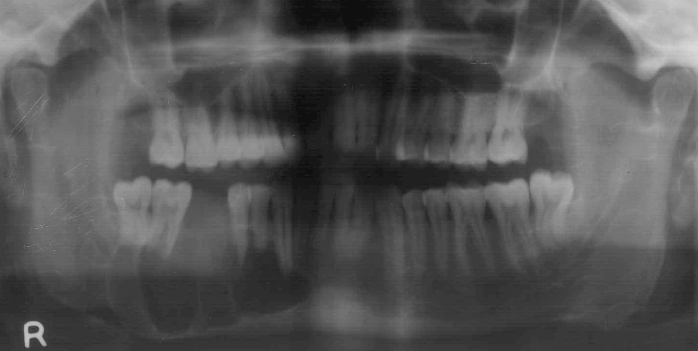
Radiographic Findings
Radiography
Unilocular or multilocular (80%) radiolucent mass with scalloped borders & thinned cortical margins.
Usually no calcifications in matrix.
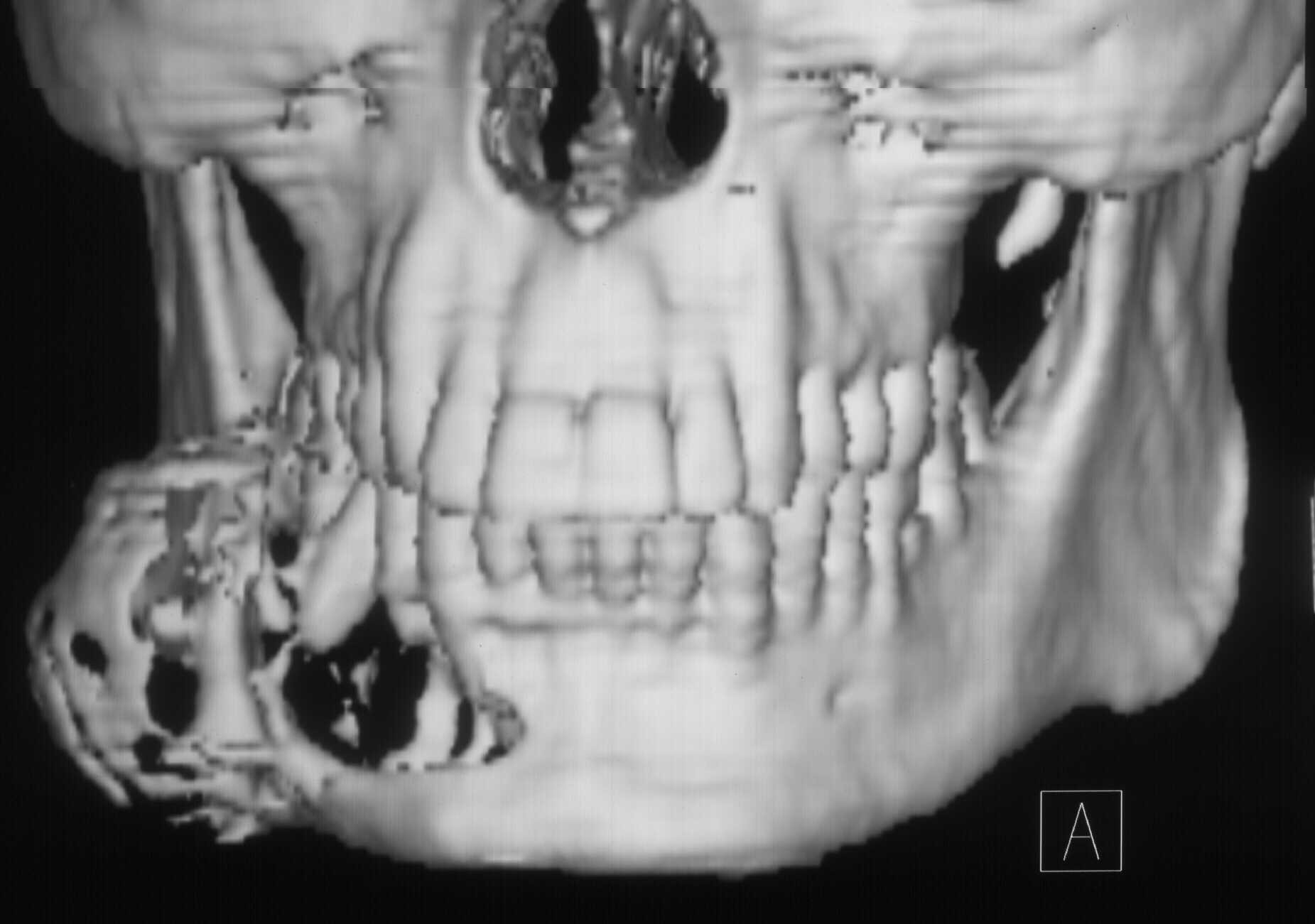
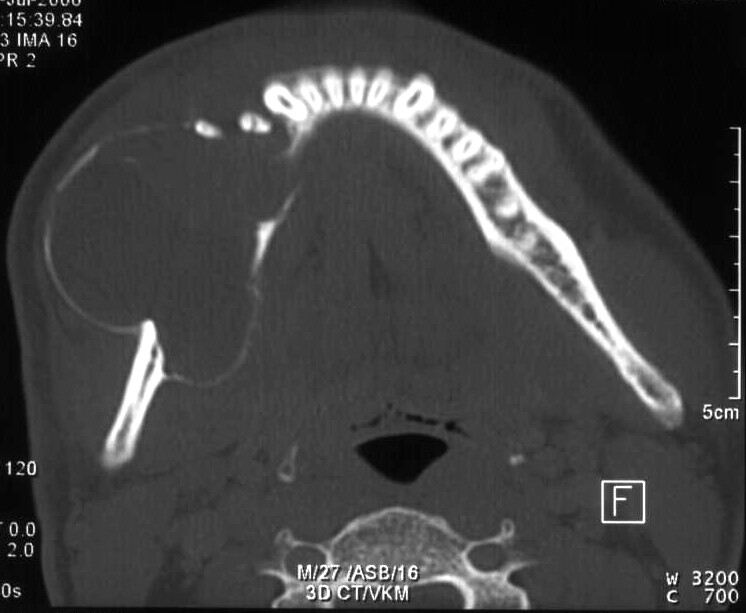
CT Findings
NECT
a. Uni- (20%) or multilocular (80%) with scalloped borders.
b. "Bubbly pattern" is typical but not pathognomonic.
c. Unerupted molar tooth association common.
d. Resorption of adjacent teeth.
e.Thinning of mandible or maxilla well-defined cortex is usually extensive. f. Low density, "osteolytic" lesion that does not mineralize its matrix.
Here is the CT of the above patient,