


Case report:
A 23 year old G1P0 lady presented to her obstetrician with history of 9 weeks of amenorrhea, lower abdominal pain and PV bleeding since one day. H/o ovulation induction with clomephene citrate is noted.An ultrasound was adviced to rule out Ectopic pregnancy .Lower abdominal tenderness is noted on clinical examination. The ultrasound examination showed twin intrauterine gestational sacs of gestational age of 8wk4d and 9wk respectively. Figure 1 Case report:
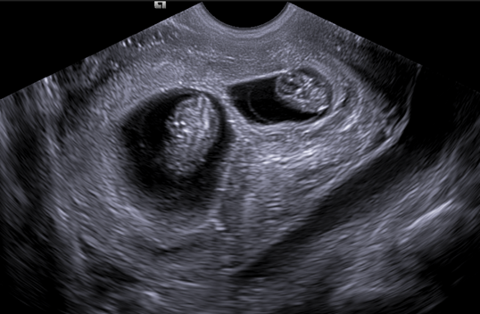
Figure 1
Ultrasound image of the saggital section of the pelvis showing two intrauterine gestational sacs with two fetal poles ,two amniotic cavities and a thick echogenic septa separating both the fetal poles. Note the free fluid with homogenous echos in posterior cul de sac.
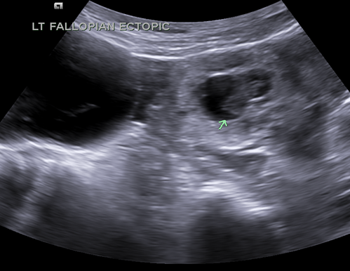
Figure 2
Thick echogenic ring like structure seen surrounding fetal pole like looking structure is seen in left adnexa. Which did not had any cardiac activity(shown with green arrow). Note the intrauterine gestation in the uterine cavity.
Figure 3
The fallopian tube held with bobs forcep contain the ectopic non viable pregnancy. Salpingotomy was performed and the intrauterine pregnancy were preserved.
Discussion.
Figure 1
Ultrasound image of the saggital section of the pelvis showing two intrauterine gestational sacs with two fetal poles ,two amniotic cavities and a thick echogenic septa separating both the fetal poles. Note the free fluid with homogenous echos in posterior cul de sac.
Figure 2
Thick echogenic ring like structure seen surrounding fetal pole like looking structure is seen in left adnexa. Which did not had any cardiac activity(shown with green arrow). Note the intrauterine gestation in the uterine cavity.
Figure 3
The fallopian tube held with bobs forcep contain the ectopic non viable pregnancy. Salpingotomy was performed and the intrauterine pregnancy were preserved.
Discussion.
Definition: Simultaneous development of a gestation within the uterine cavity and a gestation outside the uterine cavity.
Ultrasound is the most important and often only imaging modality in the imaging of ectopic pregnancy.
Demonstration of the live fetus in the adnexa is the most important diagnostic feature in the determination of the ectopic pregnancy but it is only seen in minute number of cases.
Hyperechoic ring around the gestational sac in the adnexa which on color Doppler shows ring of fire appearance. Free fluid in abdomen and pelvis with homogenous dense echos which on tapping yields blood.